

Iron inhibits activation-induced cytidine deaminase enzymatic activity and modulates immunoglobulin class switch DNA recombination
- PMID: 22556412
- PMCID: PMC3375573
- DOI: 10.1074/jbc.M112.366732
Iron inhibits activation-induced cytidine deaminase enzymatic activity and modulates immunoglobulin class switch DNA recombination
Abstract
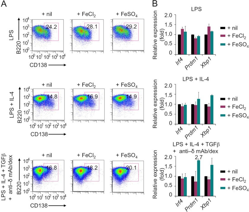
Immunoglobulin (Ig) class switch DNA recombination (CSR) and somatic hypermutation (SHM) are critical for the maturation of the antibody response. Activation-induced cytidine deaminase (AID) initiates CSR and SHM by deaminating deoxycytidines (dCs) in switch (S) and V(D)J region DNA, respectively, to generate deoxyuracils (dUs). Processing of dUs by uracil DNA glycosylase (UNG) yields abasic sites, which are excised by apurinic/apyrimidinic endonucleases, eventually generating double strand DNA breaks, the obligatory intermediates of CSR. Here, we found that the bivalent iron ion (Fe(2+), ferrous) suppressed CSR, leading to decreased number of switched B cells, decreased postrecombination Iμ-C(H) transcripts, and reduced titers of secreted class-switched IgG1, IgG3, and IgA antibodies, without alterations in critical CSR factors, such as AID, 14-3-3γ, or PTIP, or in general germline I(H)-S-C(H) transcription. Fe(2+) did not affect B cell proliferation or plasmacytoid differentiation. Rather, it inhibited AID-mediated dC deamination in a dose-dependent fashion. The inhibition of intrinsic AID enzymatic activity by Fe(2+) was specific, as shown by lack of inhibition of AID-mediated dC deamination by other bivalent metal ions, such as Zn(2+), Mn(2+), Mg(2+), or Ni(2+), and the inability of Fe(2+) to inhibit UNG-mediated dU excision. Overall, our findings have outlined a novel role of iron in modulating a B cell differentiation process that is critical to the generation of effective antibody responses to microbial pathogens and tumoral cells. They also suggest a possible role of iron in dampening AID-dependent autoimmunity and neoplastic transformation.
Figures


Similar articles
-
AID- and Ung-dependent generation of staggered double-strand DNA breaks in immunoglobulin class switch DNA recombination: a post-cleavage role for AID.Mol Immunol. 2008 Nov;46(1):45-61. doi: 10.1016/j.molimm.2008.07.003. Epub 2008 Aug 28. Mol Immunol. 2008. PMID: 18760480 Free PMC article.
-
Endonuclease G plays a role in immunoglobulin class switch DNA recombination by introducing double-strand breaks in switch regions.Mol Immunol. 2011 Jan;48(4):610-22. doi: 10.1016/j.molimm.2010.10.023. Epub 2010 Nov 26. Mol Immunol. 2011. PMID: 21111482 Free PMC article.
-
Single-strand DNA breaks in Ig class switch recombination that depend on UNG but not AID.Int Immunol. 2008 Nov;20(11):1381-93. doi: 10.1093/intimm/dxn097. Epub 2008 Sep 15. Int Immunol. 2008. PMID: 18794203
-
Opinion: uracil DNA glycosylase (UNG) plays distinct and non-canonical roles in somatic hypermutation and class switch recombination.Int Immunol. 2014 Oct;26(10):575-8. doi: 10.1093/intimm/dxu071. Epub 2014 Jul 3. Int Immunol. 2014. PMID: 24994819 Free PMC article. Review.
-
Regulation of Aicda expression and AID activity.Autoimmunity. 2013 Mar;46(2):83-101. doi: 10.3109/08916934.2012.749244. Epub 2013 Jan 17. Autoimmunity. 2013. PMID: 23181381 Free PMC article. Review.
Cited by
-
Epigenetics of the antibody response.Trends Immunol. 2013 Sep;34(9):460-70. doi: 10.1016/j.it.2013.03.006. Epub 2013 May 2. Trends Immunol. 2013. PMID: 23643790 Free PMC article. Review.
-
Influence of host iron status on Plasmodium falciparum infection.Front Pharmacol. 2014 May 6;5:84. doi: 10.3389/fphar.2014.00084. eCollection 2014. Front Pharmacol. 2014. PMID: 24834053 Free PMC article. Review.
-
Accelerated Systemic Autoimmunity in the Absence of Somatic Hypermutation in 564Igi: A Mouse Model of Systemic Lupus with Knocked-In Heavy and Light Chain Genes.Front Immunol. 2017 Sep 13;8:1094. doi: 10.3389/fimmu.2017.01094. eCollection 2017. Front Immunol. 2017. PMID: 28955333 Free PMC article.
-
Complex Interactions in Regulation of Haematopoiesis-An Unexplored Iron Mine.Genes (Basel). 2021 Aug 20;12(8):1270. doi: 10.3390/genes12081270. Genes (Basel). 2021. PMID: 34440444 Free PMC article. Review.
-
Consensus on the Key Characteristics of Immunotoxic Agents as a Basis for Hazard Identification.Environ Health Perspect. 2022 Oct;130(10):105001. doi: 10.1289/EHP10800. Epub 2022 Oct 6. Environ Health Perspect. 2022. PMID: 36201310 Free PMC article.
References
-
- Murphy K. M. (2011) in Janeway's Immunobiology (Murphy K. M., ed), 8th Ed., pp. 387–428, Garland Science, New York
-
- Odegard V. H., Schatz D. G. (2006) Targeting of somatic hypermutation. Nat. Rev. Immunol. 6, 573–583 - PubMed
-
- Casali P. (2009) in Lewin's Genes X (Krebs J. E., Goldstein E. S., Kilpatrick S. T., eds), 10th Ed., pp. 570–623, Jones & Bartlett, Sudbury, MA
-
- Goodnow C. C., Vinuesa C. G., Randall K. L., Mackay F., Brink R. (2010) Control systems and decision making for antibody production. Nat. Immunol. 11, 681–688 - PubMed
Publication types
MeSH terms
Substances
Grants and funding
LinkOut - more resources
Full Text Sources
Medical
Miscellaneous
