. 2012 Jun 15;287(25):21290-302.
doi: 10.1074/jbc.M112.357202. Epub 2012 May 3.

Activation of Wnt11 by transforming growth factor-β drives mesenchymal gene expression through non-canonical Wnt protein signaling in renal epithelial cells

Affiliations

Activation of Wnt11 by transforming growth factor-β drives mesenchymal gene expression through non-canonical Wnt protein signaling in renal epithelial cells

Peng Zhang et al. J Biol Chem. .

Abstract

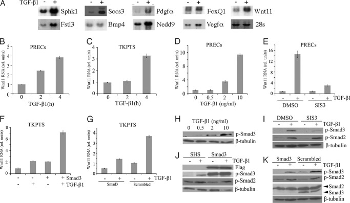
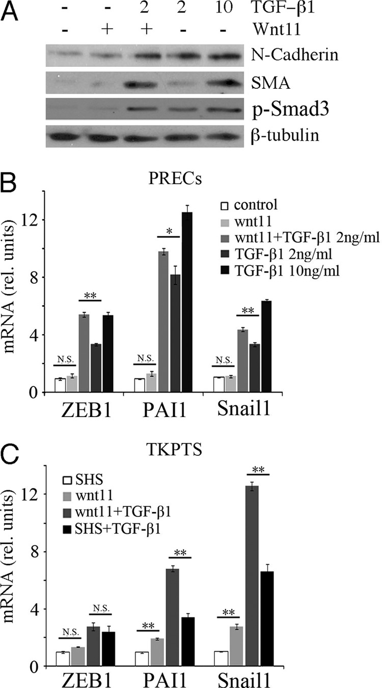
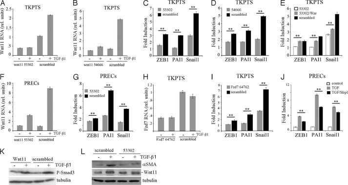
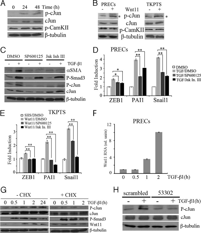
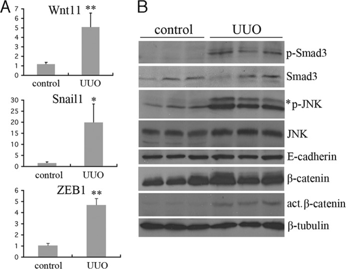
Transforming growth factor β1 (TGF-β) promotes renal interstitial fibrosis in vivo and the expression of mesenchymal genes in vitro; however, most of its direct targets in epithelial cells are still elusive. In a screen for genes directly activated by TGF-β, we found that components of the Wnt signaling pathway, especially Wnt11, were targets of activation by TGF-β and Smad3 in primary renal epithelial cells. In gain and loss of function experiments, Wnt11 mediated the actions of TGF-β through enhanced activation of mesenchymal marker genes, such as Zeb1, Snail1, Pai1, and αSMA, without affecting Smad3 phosphorylation. Inhibition of Wnt11 by receptor knockdown or treatment with Wnt inhibitors limited the effects of TGF-β on gene expression. We found no evidence that Wnt11 activated the canonical Wnt signaling pathway in renal epithelial cells; rather, the function of Wnt11 was mediated by the c-Jun N-terminal kinase (JNK) pathway. Consistent with the in vitro results, all the TGF-β, Wnt11, and JNK targets were activated in a unilateral ureteral obstruction (UUO) model of renal fibrosis in vivo. Our findings demonstrated cooperativity among the TGF-β, Wnt11, and JNK signaling pathways and suggest new targets for anti-fibrotic therapy in renal tissue.


Figures

FIGURE 1.
FIGURE 1.
Effects of TGF-β on primary renal epithelial cells. A, shown are phase contrast micrographs of PRECs treated with increasing concentrations of TGF-β for 24 h. B, shown is a Western blot of cells treated for 0, 48, and 72 h with 10 ng/ml TGF-β and probed for E-cadherin. C, shown is time and dose dependence of αSMA and N-cadherin expression with increasing amounts of TGF-β. D, shown is qRT-PCR of mesenchymal markers after 24 h of TGF-β treatment at the indicated doses. All samples were done in triplicate with error bars representing 1 S.D.
FIGURE 2.
FIGURE 2.
Activation of Wnt11 and TGF-β targets. A, shown are Northern blots of total RNA from control cells (−) or 4 h TGF-β treated cells (+) cultured in the presence of cycloheximide and probed for the indicated RNAs. B, shown are Wnt11 RNA levels in PRECs cells cultured with TGF-β for the indicated time in hours in the presence of cycloheximide. C, shown are Wnt11 RNA levels in TKPTS cells cultured with TGF-β for the indicated time in hours in the presence of cycloheximide. D, shown are Wnt11 RNA levels after 24 h with varying does of TGF-β as indicated. E, shown is Wnt11 RNA activation in response to TGF-β in the presence or absence of the Smad3 inhibitor SIS3. F, shown are Wnt11 RNA levels measured after Smad3 transfection and/or TGF-β treatment. G, shown is Wnt11 RNA activation in response to TGF-β in the presence or absence of Smad3 shRNAs or scrambled controls. H, shown is a Western blot of P-Smad3 in response to increasing doses of TGF-β. I, shown are Western blots of P-Smad3 and P-Smad2 after inhibition by SIS3 and treatment with TGF-β. J, shown is a Western blot for P-Smad3 and P-Smad2 after FLAG-Smad3 transfection or control transfection (SHS) and TGF-β treatment. K, shown is a Western blot of P-Samd3, P-Smad2, and total Smad2/3 after culture with Smad3 shRNAs and TGF-β treatment. It is noted that Smad3 proteins were shown nearly totally gone in Smad2/3 panel, whereas they were still detectable in the P-Smad3 panel. This resulted from different exposure times and different affinities for these two antibodies to their respective antigens.
FIGURE 3.
FIGURE 3.
Wnt11 increases TGF-β-dependent activation of mesenchymal genes. A, Western blots for N-cadherin, αSMA, P-Samd3, and β-tubulin from cells treated with recombinant Wnt11 and different doses of TGF-β are indicated. Note that Wnt11 increases αSMA levels at low doses of TGF-β. B, quantitative RT-PCR for genes are indicated under similar conditions as in A. C, quantitative RT-PCR for mesenchymal genes after transfection with Wnt11 expression plasmids and/or TGF-β addition. SHS, sonicated herring sperm DNA transfection control (*, p < 0.05; **, p < 0.01; N.S., not significant; Student's t test for independent variables).
FIGURE 4.
FIGURE 4.
Wnt11 is necessary for the TGF-β-dependent activation of mesenchymal genes. A, shown are Wnt11 RNA levels after culture with shRNA #53302 or a scrambled control in TKPTS cells with or without TGF-β. B, shown is a similar experiment as in A but using the Wnt11 shRNA #54666. C, shown is TGF-β induction of RNAs for the indicated genes in the presence or absence of shRNA #53302. Relative amount of RNA is compared before or after TGF-β addition and expressed as fold induction. D, shown is a similar experiment as in C but using the Wnt11 shRNA #54666. E, the induction -fold change of indicated genes by TGF-β is measured in cells cultured with shRNA 53302, with shRNA 53302 and recombinant Wnt11, or with scrambled shRNA. Note that recombinant Wnt11 increases the induction of mesenchymal genes in the presence of 53302. F, Wnt11 shRNA 53302 in PRECs reduces TGF-β-dependent Wnt11 RNA induction. G, in PRECs, Wnt11 shRNA 53302 reduced the TGF-β-mediated fold induction of mesenchymal marker genes. H, in TKPTS, inhibition of Fzd7 RNA by shRNA #64762 is independent of TGF-β. I, Fzd7 shRNA #64762 inhibits the TGF-β-mediated induction of mesenchymal marker genes. J, the Wnt secreted inhibitor Sfrp1 reduces the TGF-β-mediated induction of mesenchymal marker genes in PRECs. K, shown is a Western blot of P-Smad3 from cells cultured with Wnt11 53302 shRNAs and/or TGF-β as indicated in TKPTS. L, Western blots for αSMA cells show that inhibition of Wnt11 by shRNAs reduces the accumulation of αSMA in response to TGF-β in PRECs. Levels of Wnt11 protein induction by TGF-β are reduced by 53302 treatment, although basal levels without TGF-β are similar. *, p < 0.05; **, p < 0.01, Student's t test for independent variables.
FIGURE 5.
FIGURE 5.
Wnt11 does not mediate Smad2/3 phosphorylation or β-catenin-dependent gene activation. A, Western blots from cells overexpressing Wnt11 and treated with TGF-β show no effects of Wnt11 on P-Smad3 levels. B, the P-Smad2/3 reporter 3TP-Luc was assayed after co-transfection with Wnt11 or treatment with TGF-β. Note that Wnt11 does not increase 3TP-dependent luciferase. C, Wnt11 53302 shRNA knockdown does not affect the ability of TGF-β to activate 3TP-luc. D, a Western blot using antibodies against activated β-catenin shows no effects of Wnt11 on active β-catenin accumulation in press. E, shown is a Western blot using antibody against total β-catenin in the cytoplasmic and nuclear fractions of PRECs under Wnt11 or LiCl treatment for 24 h. Fractionation controls are β-tubulin for cytoplasma and ptip for nuclear fractions. F, Wnt11 does not activate Wisp1 or Axin, two known β-catenin target genes, as assayed by qRT-PCR in renal epithelial cells. N.S., not significant. G, cells were transfected with the β-catenin reporter TOPFLASH then treated with TGF-β or LiCl or co-transfected with Smad3/Wnt11. Only the known GSK3 kinase inhibitor LiCl significantly activated the TOPFLASH reporter. FOPFLASH was used as an internal control plasmid, and statistical significance used Student's t test for independent variables.
FIGURE 6.
FIGURE 6.
Activation of JNK signaling by TGF-β/Wnt11 activates mesenchymal marker genes. A, a Western blot for (Ser(P)-63)-c-Jun (p-cJun), total c-Jun, and P-CaMKII after treatment of PRECs for 24 or 48 h with TGF-β shows increased P- c-Jun and total c-Jun but not P-CaMKII. B, the addition of recombinant Wnt11 in PRECs or Wnt11 overexpression in TKPTS increases levels of phospho-c-Jun but not phospho-CaMKII. C, Western blots of cell lysates show that inhibition of the c-Jun kinase (JNK) by SP600125 or JNK inhibitor III reduces expression of αSMA in response to TGF-β. Note there is no affect on phospho-Smad3 levels. D, qRT-PCR of mesenchymal marker genes from samples treated with TGF-β with or without the JNK inhibitors show reduced expression of all mesenchymal markers tested upon JNK inhibition in PRECs. E, activation of mesenchymal marker gene expression in response to Wnt11 is reduced upon JNK inhibition in TKPTS, as determined by qRT-PCR. F, shown are Wnt11 RNA levels in PRECs cells cultured with TGF-β for the indicated time in hours. G, shown are Western blots for phospho-c-Jun, total c-Jun, P-Smad3, and Wnt11 with TGF-β treatment for the indicated times in the absence or in the presence of cycloheximide (CHX). Cycloheximide abolished the elevated phospho-c-Jun and Wnt11 induction upon TGF-β treatment. H, Western blots for phospho-c-Jun show reduced levels upon TGF-β treatment when cells are cultured with shRNA 53302 against Wnt11. *, p < 0.05; **, p < 0.01; Student's t test for independent variables.
FIGURE 7.
FIGURE 7.
Activation of Wnt11 and JNK in the UUO model of renal fibrosis. A, Gene expression levels were assayed by qRT-PCR for the indicated genes in control (n = 3) and 7-day UUO kidney (n = 3) RNA isolates. RNA levels are expressed as relative units with the control kidney values set at 1 (*, p < 0.05; **, p < 0.01; Student's t test for independent variables). B, shown are Western blots of kidney protein lysates from three independent control or 7-day UUO kidneys. Identical blots were probed with the indicated antibodies. Note the increased levels of P-Smad3 and P-JNK in the UUO kidney lysates.

Similar articles

Cited by

References

    1. Liu Y. (2010) New insights into epithelial-mesenchymal transition in kidney fibrosis. J Am. Soc. Nephrol. 21, 212–222 - PMC - PubMed
    1. Böttinger E. P., Bitzer M. (2002) TGF-β signaling in renal disease. J. Am. Soc. Nephrol. 13, 2600–2610 - PubMed
    1. Yang J., Liu Y. (2001) Dissection of key events in tubular epithelial to myofibroblast transition and its implications in renal interstitial fibrosis. Am. J. Pathol. 159, 1465–1475 - PMC - PubMed
    1. Zeisberg E. M., Tarnavski O., Zeisberg M., Dorfman A. L., McMullen J. R., Gustafsson E., Chandraker A., Yuan X., Pu W. T., Roberts A. B., Neilson E. G., Sayegh M. H., Izumo S., Kalluri R. (2007) Endothelial-to-mesenchymal transition contributes to cardiac fibrosis. Nat. Med. 13, 952–961 - PubMed
    1. Kopp J. B., Factor V. M., Mozes M., Nagy P., Sanderson N., Böttinger E. P., Klotman P. E., Thorgeirsson S. S. (1996) Transgenic mice with increased plasma levels of TGF-β 1 develop progressive renal disease. Lab. Invest. 74, 991–1003 - PubMed

Publication types

MeSH terms