. 2012 Jun 15;287(25):21450-60.
doi: 10.1074/jbc.M111.331249. Epub 2012 May 3.

Critical role of filamin-binding LIM protein 1 (FBLP-1)/migfilin in regulation of bone remodeling

Affiliations

Critical role of filamin-binding LIM protein 1 (FBLP-1)/migfilin in regulation of bone remodeling

Guozhi Xiao et al. J Biol Chem. .

Abstract

Bone remodeling is a complex process that must be precisely controlled to maintain a healthy life. We show here that filamin-binding LIM protein 1 (FBLP-1, also known as migfilin), a kindlin- and filamin-binding focal adhesion protein, is essential for proper control of bone remodeling. Genetic inactivation of FBLIM1 (the gene encoding FBLP-1) in mice resulted in a severe osteopenic phenotype. Primary FBLP-1 null bone marrow stromal cells (BMSCs) exhibited significantly reduced extracellular matrix adhesion and migration compared with wild type BMSCs. Loss of FBLP-1 significantly impaired the growth and survival of BMSCs in vitro and decreased the number of osteoblast (OB) progenitors in bone marrow and OB differentiation in vivo. Furthermore, the loss of FBLP-1 caused a dramatic increase of osteoclast (OCL) differentiation in vivo. The level of receptor activator of nuclear factor κB ligand (RANKL), a key regulator of OCL differentiation, was markedly increased in FBLP-1 null BMSCs. The capacity of FBLP-1 null bone marrow monocytes (BMMs) to differentiate into multinucleated OCLs in response to exogenously supplied RANKL, however, was not different from that of WT BMMs. Finally, we show that a loss of FBLP-1 promotes activating phosphorylation of ERK1/2. Inhibition of ERK1/2 activation substantially suppressed the increase of RANKL induced by the loss of FBLP-1. Our results identify FBLP-1 as a key regulator of bone homeostasis and suggest that FBLP-1 functions in this process through modulating both the intrinsic properties of OB/BMSCs (i.e., BMSC-extracellular matrix adhesion and migration, cell growth, survival, and differentiation) and the communication between OB/BMSCs and BMMs (i.e., RANKL expression) that controls osteoclastogenesis.


Figures

FIGURE 1.
FIGURE 1.
Generation of FBLIM1−/− mice. A, genomic region of interest for FBLIM1 is shown on the top. The middle is the targeting construct for FBLIM1 where the exon 7 (green box) is floxed by two LoxP sites (blue triangles, center). A neomycin cassette (Neo) flanked by two FRT sites (blue rectangles) is inserted into the seventh intron. DTA, diphtheria toxic A chain gene; Neo, neomycin resistance gene. The floxed locus for FBLIM1 after homologous recombination is shown at the bottom. B, DNA from Neo-positive ES cells was digested with restriction enzyme BamHI and analyzed by Southern blot for WT (+/+, 12.1 kb) and targeted (f/+, 8.2 kb) alleles with probe as shown in A (red rectangle).
FIGURE 2.
FIGURE 2.
Inactivation of FBLIM1 results in a severe osteopenic phenotype in mice. A, three-dimensional reconstruction from microcomputerized tomography scan of femurs from 3-month-old male FBLIM1−/− mice and their WT male littermates. B–F, quantitative analysis of bone volume/tissue volume (BV/TV), trabecular number (Tb.N), trabecular thickness (Tb.Th), trabecular space (Tb.Sp), and cortical thickness (Cort. Th). *, p < 0.05 (versus WT); n = 4.
FIGURE 3.
FIGURE 3.
Loss of FBLP-1 compromises BMSC-ECM adhesion and migration. A–D, primary BMSCs isolated from WT (A and B) and FBLIM1−/− (C and D) mice were plated on collage I-coated coverslips and stained with monoclonal anti-FBLP-1 antibody (A and C) or phalloidin (B and D). Bar, 10 μm. E, primary BMSCs isolated from WT and FBLIM1−/− mice were analyzed by Western blotting with antibodies recognizing FBLP-1 or tubulin (as a loading control). F and G, BMSC-ECM adhesion. Primary BMSCs isolated from WT and FBLIM1−/− mice were plated in type I collagen-coated (F) or fibronectin-coated (G) 96-well plates and incubated at 37 °C for 15 min. Cell-ECM adhesion was analyzed using a quantitative centrifugal force assay as described under “Experimental Procedures.” Adhesion of FBLIM1−/− BMSCs was compared with that of WT BMSCs (normalized to 100%). The bars represent the means ± S.D. from three independent experiments. *, p < 0.05 (versus WT). H and I, MC4 cell-ECM adhesion. MC4 cells were transfected with FBLP-1 or control siRNA. The FBLP-1 siRNA and control transfectants were analyzed by Western blotting with antibodies recognizing FBLP-1 or tubulin (H) or a quantitative centrifugal force cell-ECM adhesion assay (I). Adhesion of FBLP-1 knockdown MC4 cells was compared with that of control MC4 cells (normalized to 100%). The bars in I represent the means ± S.D. from three independent experiments. *, p < 0.05 (versus control). J, cell migration was assessed with a scratch assay as described under “Experimental Procedures.” The distances traveled by FBLIM1−/− BMSCs at the acellular fronts were measured 6.5 h after wounding and compared with those of WT BMSCs (normalized to 100%). The bars represent the means ± S.D. from three independent experiments. *, p < 0.05 (versus WT).
FIGURE 4.
FIGURE 4.
FBLP-1 ablation compromises the formation of OB progenitors in bone marrow and OB differentiation in vivo. A and B, CFU-F assay. 1 × 10⁶ bone marrow-nucleated cells from WT and FBLIM1−/− mice were cultured for 10 days, and CFU-Fs were quantified as described under “Experimental Procedures.” The bars in B represent the means ± S.D. from three experiments. C and D, CFU-OB assay. 1 × 10⁶ bone marrow-nucleated cells from WT and FBLIM1−/− mice were cultured in OB differentiation medium for 21 days, and the numbers of CFU-OB colonies were quantified as described under “Experimental Procedures.” The bars in D represent the means ± S.D. from four experiments. *, p < 0.05 (versus WT). E, BMSC growth. WT and FBLIM1−/− BMSCs were seeded at a density of 1 × 10⁴/well in 96-well plates in proliferation medium. MTS assays were performed on days 0, 2, 4, and 6 (d0, d2, d4, and d6, respectively) as indicated. *, p < 0.05 (versus WT). F and G, TUNEL staining. Apoptosis of WT and FBLIM1−/− BMSCs was analyzed by TUNEL staining as described under “Experimental Procedures.” The arrows in F indicate TUNEL-positive cells. The bars in G represent the means ± S.D. from five experiments. *, p < 0.05 (versus WT). H and I, Osx staining. Five-μm tibial sections from 3-month-old WT and FBLIM1−/− male mice were immunohistochemically stained with anti-Osx antibody or control IgG as indicated in H. The arrows in H indicate the nuclei of Osx-positive OBs that were stained brown. Osx-negative cells were stained blue. Original magnification was ×100 (top) and ×200 (bottom). I, the numbers of Osx-positive cells (i.e., differentiating and differentiated OBs) located on trabecular bone surfaces were counted and normalized to trabecular bone perimeter (Osx (+) Ob.nb/BPm) using an Image Pro Plus 6.2 software. *, p < 0.05 (versus WT); n = 4. J, real time RT-PCR. Tibiae from 2-month-old WT and FBLIM1−/− mice (n = 4) were harvested for RNA isolation. Total RNA was used for quantitative real time PCR using specific primers for Osx mRNAs, which were normalized to GAPDH mRNA. *, p < 0.05 (versus WT); n = 4.
FIGURE 5.
FIGURE 5.
FBLP-1 deficiency dramatically increases OCL differentiation in vivo. A, TRAP staining. Tibial sections of 3-month-old WT and FBLIM1−/− male mice were stained for TRAP activity for 30 min at 37 °C. B, TRAP activity in metaphyseal regions of tibias is shown. The arrows indicate TRAP-positive MNCs on trabecular surfaces. C, OCL surface/bone surface (Oc.S/BS) and OCL number/bone perimeter (Oc.Nb/BPm) in both primary and secondary spongiosa of tibiae in A were measured as described previously (33). *, p < 0.05 (versus WT); n = 4
FIGURE 6.
FIGURE 6.
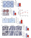
Loss of FBLP-1 promotes OCL differentiation via up-regulation of RANKL expression in OBs/BMSCs. A and B, TRAP staining. WT and FBLIM1−/− BMM differentiation were induced with M-CSF (10 ng/ml) and RANKL (50 ng/ml) for 7 days, followed by TRAP staining (A). TRAP-positive MNCs (three or more nuclei) per well were scored (B). C, BMSC-BMM coculture. Primary BMSCs and BMMs from 2-month-old WT and FBLIM1−/− mice were cocultured in the absence of exogenously supplied RANKL for 10 days, followed by TRAP staining. D and E, real time RT-PCR and Western blot. Primary BMSCs from 2-month-old WT and FBLIM1−/− mice were seeded at a density of 5 × 10⁴/cm2 in 35-mm dish in proliferation medium for 24 h and harvested for RNA and protein isolation. Total RNA was used for quantitative real time PCR (D) using specific primers for Rankl and Opg mRNAs, which were normalized to GAPDH mRNA. Whole cell extracts were used for Western blot with antibodies as specified in the figure (E). F, immunohistochemical staining. Five-μm tibial sections from 3-month-old WT and FBLIM1−/− male mice were immunohistochemically stained with anti-RANKL antibody or control IgG as indicated in the figure.
FIGURE 7.
FIGURE 7.
Loss of FBLP-1 increases RANKL expression via promoting ERK1/2 activation in BMSCs. A, loss of FBLP-1 promotes activating phosphorylation of ERK1/2. Primary BMSCs from 2-month-old WT and FBLIM1−/− mice were seeded at a density of 5 × 10⁴/cm2 in 35-mm dish in proliferation medium for 24 h and harvested for protein isolation and Western blot with antibodies against phospho-ERK1/2 (Thr-202/Tyr-204), ERK1/2, phosphor-AKT (Ser-473), AKT, and β-actin as indicated in the figure. B, inhibition of ERK1/2 activation suppresses FBLP-1 deficiency induced RANKL expression. Primary BMSCs from 2-month-old FBLIM1−/− mice were seeded at a density of 5 × 10⁴/cm2 in 35-mm dish in proliferation medium for 24 h and treated with and without U0126 (5 μm) for 24 h, followed by Western blotting with antibodies against RANKL, phospho-ERK1/2 (Thr-202/Tyr-204), ERK1/2, and β-actin as indicated in the figure.

References

    1. Ash P., Loutit J. F., Townsend K. M. (1980) Osteoclasts derived from haematopoietic stem cells. Nature 283, 669–670 - PubMed
    1. Lacey D. L., Timms E., Tan H. L., Kelley M. J., Dunstan C. R., Burgess T., Elliott R., Colombero A., Elliott G., Scully S., Hsu H., Sullivan J., Hawkins N., Davy E., Capparelli C., Eli A., Qian Y. X., Kaufman S., Sarosi I., Shalhoub V., Senaldi G., Guo J., Delaney J., Boyle W. J. (1998) Osteoprotegerin ligand is a cytokine that regulates osteoclast differentiation and activation. Cell 93, 165–176 - PubMed
    1. Yasuda H., Shima N., Nakagawa N., Yamaguchi K., Kinosaki M., Mochizuki S., Tomoyasu A., Yano K., Goto M., Murakami A., Tsuda E., Morinaga T., Higashio K., Udagawa N., Takahashi N., Suda T. (1998) Osteoclast differentiation factor is a ligand for osteoprotegerin/osteoclastogenesis-inhibitory factor and is identical to TRANCE/RANKL. Proc. Natl. Acad. Sci. U.S.A. 95, 3597–3602 - PMC - PubMed
    1. Simonet W. S., Lacey D. L., Dunstan C. R., Kelley M., Chang M. S., Lüthy R., Nguyen H. Q., Wooden S., Bennett L., Boone T., Shimamoto G., DeRose M., Elliott R., Colombero A., Tan H. L., Trail G., Sullivan J., Davy E., Bucay N., Renshaw-Gegg L., Hughes T. M., Hill D., Pattison W., Campbell P., Sander S., Van G., Tarpley J., Derby P., Lee R., Boyle W. J. (1997) Osteoprotegerin. A novel secreted protein involved in the regulation of bone density. Cell 89, 309–319 - PubMed
    1. O'Brien C. A. (2010) Control of RANKL gene expression. Bone 46, 911–919 - PMC - PubMed

Publication types

MeSH terms

Substances