. 2012 Jun 22;287(26):21950-6.
doi: 10.1074/jbc.M112.350389. Epub 2012 May 3.

Drosophila heparan sulfate, a novel design

Affiliations

Drosophila heparan sulfate, a novel design

Marion Kusche-Gullberg et al. J Biol Chem. .

Abstract

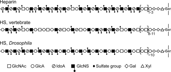
Heparan sulfate (HS) proteoglycans play critical roles in a wide variety of biological processes such as growth factor signaling, cell adhesion, wound healing, and tumor metastasis. Functionally important interactions between HS and a variety of proteins depend on specific structural features within the HS chains. The fruit fly (Drosophila melanogaster) is frequently applied as a model organism to study HS function in development. Previous structural studies of Drosophila HS have been restricted to disaccharide composition, without regard to the arrangement of saccharide domains typically found in vertebrate HS. Here, we biochemically characterized Drosophila HS by selective depolymerization with nitrous acid. Analysis of the generated saccharide products revealed a novel HS design, involving a peripheral, extended, presumably single, N-sulfated domain linked to an N-acetylated sequence contiguous with the linkage to core protein. The N-sulfated domain may be envisaged as a heparin structure of unusually low O-sulfate content.


Figures

FIGURE 1.
FIGURE 1.
Structure of HS chain and approaches for analysis. A, the structural domains (NS-, NA/NS-, and NA-domains) are defined with regard to the distribution of GlcNS and GlcNAc units. B, schematic representation of methods used to elucidate HS domain structure. For further information, see “Results” and “Experimental Procedures.” The symbols used are defined below the scheme. GlcA, GlcUA; IdoA, IdoUA.
FIGURE 2.
FIGURE 2.
Gel chromatography of 3H-labeled saccharides obtained after deaminative cleavage of HS. HS samples from Drosophila or pig intestinal mucosa were deaminated and radiolabeled according to Protocol A (A and B) or Protocol B (C and D) (see “Experimental Procedures,” Fig. 1, and supplemental Fig. S1), and the products were analyzed by Bio-Gel P-10 gel chromatography. The numbers above each peak in A indicate oligosaccharide size.
FIGURE 3.
FIGURE 3.
High-voltage paper electrophoresis of 3H-labeled disaccharides. 3H-Labeled disaccharides isolated by gel chromatography (Sephadex G-15) after deamination and radiolabeling of Drosophila HS according to Protocol A (A) or Protocol B (B) (see “Experimental Procedures,” Fig. 1, and supplemental Fig. S1) were fractionated by high-voltage electrophoresis on Whatman No. 3MM paper at pH 5.3. After drying, papers were cut into 1-cm segments and analyzed for radioactivity. Arrows indicate the migration positions of non-sulfated (1), mono-O-sulfated (2), and di-O-sulfated (3) HexUA-aManR disaccharide standards.
FIGURE 4.
FIGURE 4.
Anion-exchange HPLC of 3H-labeled disaccharides. Disaccharides generated by deamination/radiolabeling according to Protocol A (A) or Protocol B (B) were analyzed by anion-exchange HPLC on a Partisil 10 SAX column. C, end-labeled, N-sulfated oligosaccharides obtained through Protocol B were deaminated at pH 1.5, and the released labeled disaccharide was isolated and subjected to anion-exchange HPLC. (Note that the [3H]aManR residues here represent a GlcNAc unit in the intact HS chains; the corresponding reaction is illustrated to the right.) The dashed line in A refers to a sample digested with liver β-glucuronidase/α-iduronidase prior to chromatography. Arrows indicate the elution positions of disaccharide standards (with the corresponding disaccharide units in the intact HS chain shown in parentheses): 1,2, GlcUA-aManR, IdoUA-aManR (-GlcUA/IdoUA-GlcNAc/NS-), aManR, and aManR6S (generated by β-glucuronidase/α-iduronidase digestion of disaccharides) (peaks not individually identified); 3, GlcUA-aManR6S (-GlcUA-GlcNS6S-); 4, IdoUA-aManR6S (-IdoUA-GlcNS6S-); 5, IdoUA2S-aManR (-IdoUA2S-GlcNS-); 6, IdoUA2S-aManR6S (-IdoUA2S-GlcNS6S-). Note that only the aManR residues indicated by open triangles were radiolabeled (derived from GlcNAc residues in the intact polysaccharide).
FIGURE 5.
FIGURE 5.
Schematic presentation of HS/heparin chains. GlcA, GlcUA; IdoA, IdoUA.

References

    1. Feta A., Do A. T., Rentzsch F., Technau U., Kusche-Gullberg M. (2009) Molecular analysis of heparan sulfate biosynthetic enzyme machinery and characterization of heparan sulfate structure in Nematostella vectensis. Biochem. J. 419, 585–593 - PubMed
    1. Medeiros G. F., Mendes A., Castro R. A., Baú E. C., Nader H. B., Dietrich C. P. (2000) Distribution of sulfated glycosaminoglycans in the animal kingdom: widespread occurrence of heparin-like compounds in invertebrates. Biochim. Biophys. Acta 1475, 287–294 - PubMed
    1. Bernfield M., Götte M., Park P. W., Reizes O., Fitzgerald M. L., Lincecum J., Zako M. (1999) Functions of cell surface heparan sulfate proteoglycans. Annu. Rev. Biochem. 68, 729–777 - PubMed
    1. Bishop J. R., Schuksz M., Esko J. D. (2007) Heparan sulfate proteoglycans fine-tune mammalian physiology. Nature 446, 1030–1037 - PubMed
    1. Bülow H. E., Hobert O. (2006) The molecular diversity of glycosaminoglycans shapes animal development. Annu. Rev. Cell Dev. Biol. 22, 375–407 - PubMed

Publication types

MeSH terms

LinkOut - more resources