. 2012 Jun 15;287(25):21501-8.
doi: 10.1074/jbc.M112.341560. Epub 2012 May 2.

Crystal structure of elongator subcomplex Elp4-6

Affiliations

Crystal structure of elongator subcomplex Elp4-6

Zhijie Lin et al. J Biol Chem. .

Abstract

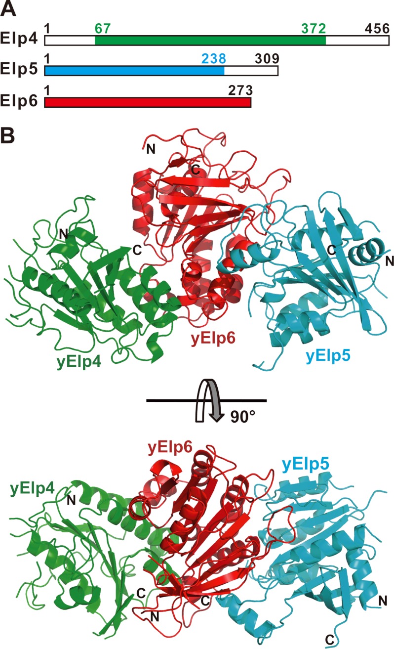
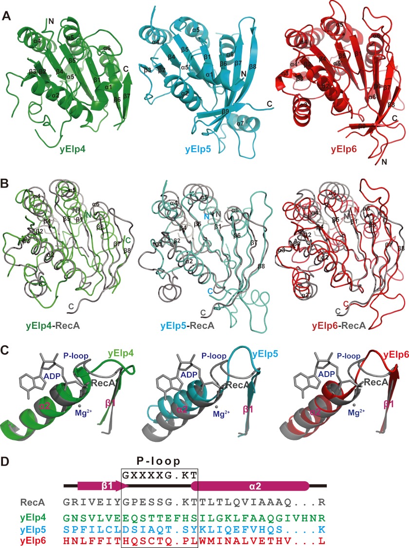
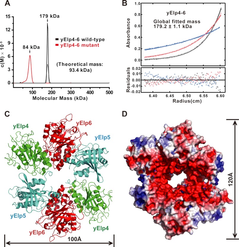
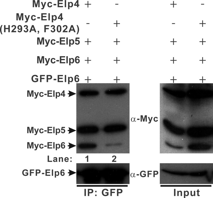
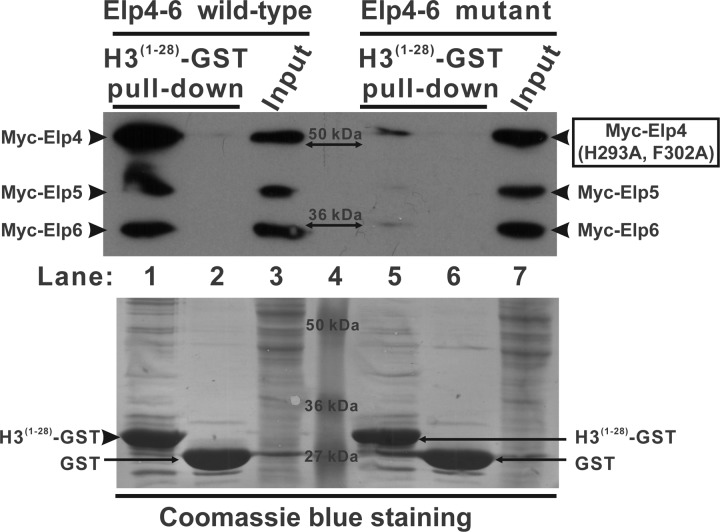
Elongator is a multiprotein complex composed of two subcomplexes, Elp1-3 and Elp4-6. Elongator is highly conserved between yeast and humans and plays an important role in RNA polymerase II-mediated transcriptional elongation and many other processes, including cytoskeleton organization, exocytosis, and tRNA modification. Here, we determined the crystal structure of the Elp4-6 subcomplex of yeast. The overall structure of Elp4-6 revealed that Elp6 acts as a bridge to assemble Elp4 and Elp5. Detailed structural and sequence analyses revealed that each subunit in the Elp4-6 subcomplex forms a RecA-ATPase-like fold, although it lacks the key sequence signature of ATPases. Site-directed mutagenesis and biochemical analyses indicated that the Elp4-6 subcomplex can assemble into a hexameric ring-shaped structure in vitro and in vivo. Furthermore, GST pulldown assays showed that the ring-shaped assembly of the Elp4-6 subcomplex is important for its specific histone H3 binding. Our results may shed light on the substrate recognition and assembly of the holo-Elongator complex.


Figures

FIGURE 1.
FIGURE 1.
Crystal structure of the yeast Elp4–6 subcomplex. A, schematic representation of Elp4, Elp5, and Elp6. The gene fragments of the tripartite complex used for structural determination in this study are as follows: yElp4 (residues 67–372) colored in green, yElp5 (residues 1–238) in cyan, and yElp6 (residues 1–273) in red. B, a schematic representation of the structure of the yElp4–6 subcomplex. yElp4, yElp5, and yElp6 are colored as in A. Shown are the side view (upper panel) and top view (lower panel). The N and C termini of these three proteins are labeled.
FIGURE 2.
FIGURE 2.
yElp4, yElp5, and yElp6 all adopt similar RecA-ATPase-like folds. A, a cartoon representation of each subunit of the yElp4–6 complex. yElp4, yElp5, and yElp6 are shown in green, cyan, and red, respectively. B, the superimposed structures of yElp4 (green), yElp5 (cyan), and yElp6 (red) with RecA (gray, Protein Data Bank code 2REB). The conserved strands and helices are marked. C, detailed structural comparison of the P-loop region between RecA (gray, ADP+Mg2+-binding form, Protein Data Bank code 1XMV) and yElp4 (green), yElp5 (cyan), or yElp6 (red). D, structure-based sequence alignment of the potential P-loop region of yElp4, yElp5, and yElp6 with that of E. coli RecA. The P-loop region is marked with a black box. The consensus sequence GXXXXGKT (X is any residue) is the Walker motif A.
FIGURE 3.
FIGURE 3.
The hexamer of the yElp4–6 subcomplex. A, SV analysis of the yElp4–6 subcomplex proteins. The wild-type tripartite yElp4–6 complex is shown with black lines, and the tripartite yElp4–6 mutant complex containing the two point mutations at H293A and F302A in yElp4 is shown with the red line. Solid and dotted lines represent protein concentrations of ∼17.6 and 8.8 μm, respectively. B, a representative SE analysis of the tripartite yElp4–6 complex derived from a global fit revealed a molecular mass of ∼179.2 ± 1.1 kDa, indicating that the wild-type yElp4–6 subcomplex assembles into a hexamer (dimer of heterotrimers). C, ribbon representation of the hexamer of the yElp4–6 subcomplex. yElp4, yElp5, and yElp6 are shown in green, cyan, and red, respectively. D, the solvent-accessible electrostatic surface representation of the hexamer of the yElp4–6 subcomplex. The surfaces are colored according to the electrostatic potential, ranging from deep blue (positive charge, +5 kT/e) to red (negative charge, −5 kT/e). The electrostatic potentials were calculated using ABPS tools (51) with its default settings.
FIGURE 4.
FIGURE 4.
The Elp4–6 subcomplex dimerizes in vivo. Extracts were prepared from HEK293T cells transfected with various combinations of plasmids as indicated, immunoprecipitated with agarose conjugated anti-GFP and subsequently immunoblotted with α-Myc (upper panels) or α-GFP (lower panels) as indicated. The left panels show the immunoprecipitation (IP) results (lane 1, wild-type Elp4–6 complex; lane 2, mutant Elp4–6 complex), and the right panels represent 2% of the input material for each immunoprecipitation. The mobilities of Myc-Elp4, Myc-Elp4 (H293A, F302A), Myc-Elp5, Myc-Elp6, and GFP-Elp6 are indicated in the left margin.
FIGURE 5.
FIGURE 5.
Interaction of the Elp4–6 subcomplex with histone H3. H3(1–28)-GST fusion proteins (lanes 1 and 5) or GST alone (lanes 2 and 6) were incubated with the extracts of HEK293T cells transfected with the wild-type or the mutant Elp4–6 complex. Lanes 3 and 7 represent 4% of the input material for the corresponding pulldown. The PVDF membrane was immunoblotted with α-Myc (upper panel) and subsequently stained with Coomassie Blue (lower panel). The mobilities of Myc-Elp4, Myc-Elp4 (H293A, F302A), Myc-Elp5, Myc-Elp6, and the GST proteins are indicated in the margins. The molecular mass of each protein marker is indicated in lane 4.

Similar articles

Cited by

References

    1. Shilatifard A., Conaway R. C., Conaway J. W. (2003) The RNA polymerase II elongation complex. Annu. Rev. Biochem. 72, 693–715 - PubMed
    1. Selth L. A., Sigurdsson S., Svejstrup J. Q. (2010) Transcript elongation by RNA polymerase II. Annu. Rev. Biochem. 79, 271–293 - PubMed
    1. Li B., Carey M., Workman J. L. (2007) The role of chromatin during transcription. Cell 128, 707–719 - PubMed
    1. Clapier C. R., Cairns B. R. (2009) The biology of chromatin remodeling complexes. Annu. Rev. Biochem. 78, 273–304 - PubMed
    1. Bannister A. J., Kouzarides T. (2011) Regulation of chromatin by histone modifications. Cell Res. 21, 381–395 - PMC - PubMed

Publication types

MeSH terms

Substances

Associated data