. 2011 Dec;8(4):259-266.
doi: 10.1007/s10388-011-0291-7. Epub 2011 Sep 10.

Usefulness of blood supply visualization by indocyanine green fluorescence for reconstruction during esophagectomy

Usefulness of blood supply visualization by indocyanine green fluorescence for reconstruction during esophagectomy

Yutaka Shimada et al. Esophagus. 2011 Dec.

Abstract

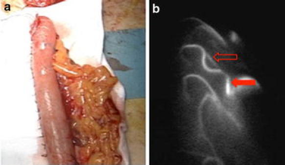
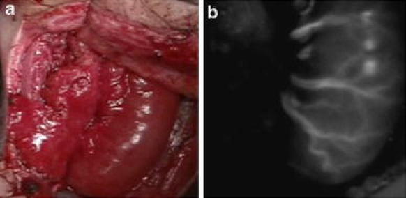
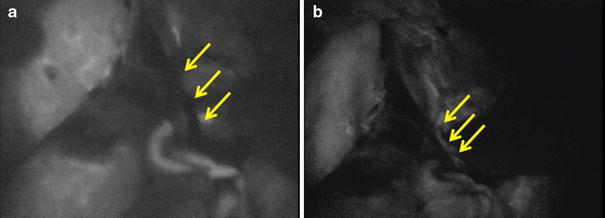
BACKGROUND: Adequate blood supply for the reconstructed organ is important for safe esophagogastric anastomosis during esophagectomy. Recently, indocyanine green (ICG) has been used for visualization of the blood supply when anastomosis is performed in vascular surgery. To visualize the blood supply for reconstruction, we employed ICG fluorescence during esophagectomy. METHODS: From August 2008, 40 patients received cervical or thoracic esophagectomy. They consisted of 33 patients having esophagectomy for thoracic esophageal cancer, 3 being treated for cervical esophageal cancer, and 4 with double cancer of the thoracic and cervical regions. Before and after pulling up the reconstructed organ, 2.5 mg of ICG was injected as a bolus. Then ICG fluorescence was detected by a camera and recorded. RESULTS: ICG fluorescence was easily detected in all patients at 1 min after injection. The vascular network was well visualized in the gastric wall, colonic grafts, and free jejunal grafts. In five patients, we also performed anastomosis between the short gastric vein and the external cervical vein or superficial cervical vein. The intraoperative and postoperative course of all patients was uneventful apart from three anastomotic leakages. CONCLUSIONS: ICG fluorescence can be employed to evaluate the blood supply to reconstructed organs and can be useful in selecting the patients who do not need additional vessel anastomosis. However, anastomotic leakage was not reduced, so the microcirculation detected by ICG fluorescence did not necessarily provide appropriate blood supply for a viable anastomosis.


Figures

Fig. 1
Fig. 1
Near-infrared camera system
Fig. 2
Fig. 2
ICG fluorescence image of a gastric tube before anastomosis. Blood flow in the arteries and veins is well visualized. a The gastric tube. b ICG fluorescence image. A closed arrow indicates an artery and an open arrow indicates a vein
Fig. 3
Fig. 3
Microvessels in the gastric wall are well visualized about 2 min after ICG injection. a Image obtained under normal light. b Blood vessels and microcirculation of the gastric tube. The forceps indicates a small blood vessel in the gastric wall and closed arrow indicates microcirculation
Fig. 4
Fig. 4
ICG fluorescence image of a free jejunal graft. a Edematous graft with repeated vascular anastomosis. b Although the graft is edematous, ICG fluorescence can visualize blood flow
Fig. 5
Fig. 5
Before and after re-anastomosis between the short gastric vein and cervical vein. a ICG fluorescence shows no blood flow in the anastomosed vessel. b ICG fluorescence shows blood flow in the vessel after repeat anastomosis
Fig. 6
Fig. 6
ICG fluorescence images of a colonic graft. a The ileo-colic vessels. b There is good blood flow from the middle colic vessels

References

    1. Alanezi K, Urschel JD. Mortality secondary to esophageal anastomotic leak. Ann Thorac Cardiovasc Surg. 2004;10:71–75. - PubMed
    1. Udagawa H, Akiyama H. Surgical treatment of esophageal cancer: Tokyo experience of the three-field technique. Dis Esophagus. 2001;14:110–114. doi: 10.1046/j.1442-2050.2001.00166.x. - DOI - PubMed
    1. Ando N, Ozawa S, Kitagawa Y, Shinozawa Y, Kitajima M. Improvement in the results of surgical treatment of advanced squamous esophageal carcinoma during 15 consecutive years. Ann Surg. 2000;232:225–232. doi: 10.1097/00000658-200008000-00013. - DOI - PMC - PubMed
    1. Tachimori Y, Kanamori N, Uemura N, Hokamura N, Igaki H, Kato H. Salvage esophagectomy after high-dose chemoradiotherapy for esophageal squamous cell carcinoma. J Thorac Cardiovasc Surg. 2009;137:49–54. doi: 10.1016/j.jtcvs.2008.05.016. - DOI - PubMed
    1. Korst R, Port JL, Lee PC, Altorki NK. Intrathoracic manifestations of cervical anastomotic leaks after transthoracic esophagectomy for carcinoma. Ann Thorac Surg. 2005;80:1185–1190. doi: 10.1016/j.athoracsur.2005.04.020. - DOI - PubMed

LinkOut - more resources