

Paragangliomas and paraganglioma syndromes
- PMID: 22558053
- PMCID: PMC3341580
- DOI: 10.3205/cto000076
Paragangliomas and paraganglioma syndromes
Abstract
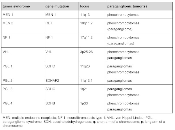
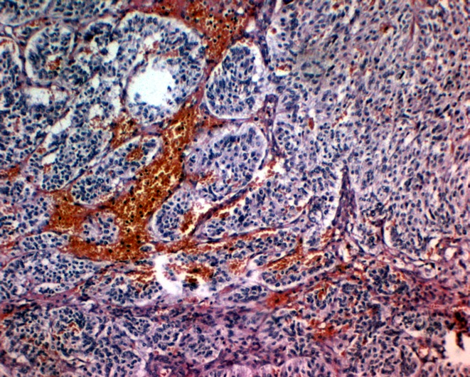
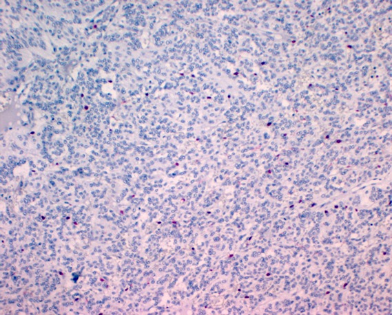
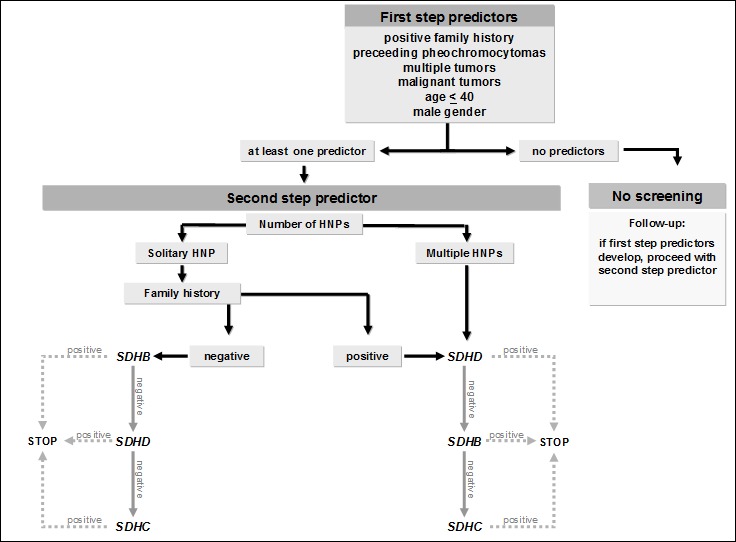
Paragangliomas are rare tumors of neural crest origin. They are benign in the majority of cases and are characterized by a strong vascularisation.In the head and neck region they most commonly occur as carotid body tumors. Jugulotympanic and especially vagal paragangliomas are seen less frequently. Complete surgical resection represents the only curative treatment option even though resection of locally advanced tumors regularly results in lesions of the lower cranial nerves and major vessels. Appoximately 30% of all head and neck paragangliomas (HNPs) are hereditary and associated with different tumor syndromes. The paraganglioma syndromes 1, 3 and 4 (PGL 1, 3 and 4) make up the majority of those familial cases. PGL 1 is associated with mutations of the succinate dehydrogenase subunit D (SDHD) gene, PGL 3 is caused by SDHC and PGL 4 by SDHB gene mutations. Multiple HNPs and the occurance of HNPs together with pheochromocytomas are seen in SDHD as well as SDHB mutation carriers. In patients with SDHB mutations the risk for the development of malignant paraganglial tumors is significantly higher compared to SDHC and SDHD patients as well as patients with sporadic tumors. SDHC mutation carriers almost exclusively present with benign HNP that are unifocal in the majority of cases. The role of transmission is autosomal dominant for all three symptoms. Interestingly, there is a "parent-of-origin-dependent-inheritance" in subjects with SDHD gene mutations. This means that the disease phenotype may only become present if the mutation is inherited through the paternal line. We recommend screening for mutations of the genes SDHB, SDHC and SDHD in patients with HNPs. Certain clinical parameters can help to set up the order in which the three genes should be tested.
Keywords: glomus tumor; paraganglioma; paraganglioma syndrome; pheochromocytoma; rare diseases.
Figures
References
-
- Boedeker CC, Ridder GJ, Neumann HPH, Maier W, Schipper J. Diagnostik, Therapie und Behandlungsergebnisse zervikaler Paragangliome. [Diagnosis and management of cervical paragangliomas: the Freiburg experience]. Laryngo-Rhino-Otol. 2004;83:585–592. doi: 10.1055/s-2004-814466. (Ger). Available from: http://dx.doi.org/10.1055/s-2004-814466. - DOI - PubMed
-
- Schipper J, Boedeker CC, Maier W, Neumann HPH. Paragangliome im Kopf-/Halsbereich Teil 1: Systematik und Diagnostik. [Paragangliomas in the head-/neck region. I: Classification and diagnosis]. HNO. 2004;52:569–575. (Ger). - PubMed
-
- Persky MS, Setton A, Niimi Y, Hartman J, Frank D, Berenstein A. Combined endovascular and surgical treatment of head and neck paragangliomas - a team approach. Head Neck. 2002;24:423–431. doi: 10.1002/hed.10068. Available from: http://dx.doi.org/10.1002/hed.10068. - DOI - PubMed
-
- Boedeker CC, Ridder GJ, Schipper J. Paragangliomas of the head and neck: diagnosis and treatment. Fam Cancer. 2005;4:55–59. doi: 10.1007/s10689-004-2154-z. Available from: http://dx.doi.org/10.1007/s10689-004-2154-z. - DOI - PubMed
-
- Boedeker CC, Neumann HPH, Offergeld C, Maier W, Falcioni M, Berlis A, Schipper J. Clinical features of paraganglioma syndromes. Skull Base. 2009;19:17–25. doi: 10.1055/s-0028-1103123. Available from: http://dx.doi.org/10.1055/s-0028-1103123. - DOI - PMC - PubMed
LinkOut - more resources
Full Text Sources
