. 2011:10:Doc06.
doi: 10.3205/cto000079. Epub 2012 Apr 26.

Molecular biology of hearing

Affiliations

Molecular biology of hearing

Timo Stöver et al. GMS Curr Top Otorhinolaryngol Head Neck Surg. 2011.

Abstract

THE INNER EAR IS OUR MOST SENSITIVE SENSORY ORGAN AND CAN BE SUBDIVIDED INTO THREE FUNCTIONAL UNITS: organ of Corti, stria vascularis and spiral ganglion. The appropriate stimulus for the organ of hearing is sound, which travels through the external auditory canal to the middle ear where it is transmitted to the inner ear. The inner ear houses the hair cells, the sensory cells of hearing. The inner hair cells are capable of mechanotransduction, the transformation of mechanical force into an electrical signal, which is the basic principle of hearing. The stria vascularis generates the endocochlear potential and maintains the ionic homeostasis of the endolymph. The dendrites of the spiral ganglion form synaptic contacts with the hair cells. The spiral ganglion is composed of neurons that transmit the electrical signals from the cochlea to the central nervous system. In recent years there has been significant progress in research on the molecular basis of hearing. An increasing number of genes and proteins related to hearing are being identified and characterized. The growing knowledge of these genes contributes not only to greater appreciation of the mechanism of hearing but also to a deeper understanding of the molecular basis of hereditary hearing loss. This basic research is a prerequisite for the development of molecular diagnostics and novel therapies for hearing loss.

Keywords: cochlea; deafness; hair cell; inner ear; organ of Corti; spiral ganglion.


Figures

Table 1
Table 1. Genes associated with hearing loss [18]. Listed below is a selection of genes, the proteins for which they code, the available mouse mutants and the form of hearing loss associated with each gene mutation. All genes listed are expressed in hair bundles and are essential for the development and/or function of the hair bundles.
Table 2
Table 2. Currently known genes that, in the case of mutation, alter K+ homeostasis [4]. The gene, the coded protein, its location and function are listed, as well as the associated type of hearing loss.
Figure 1
Figure 1. Schematic representation of the organ of Corti. The figure shows the different cell types and extracellular structures in the organ of Corti. Abbreviations: TM (tectorial membrane), OHC (outer hair cells), IHC (inner hair cells), HB (hair bundle), SC (supporting cells), CT (Corti’s tunnel).
Figure 2
Figure 2. Anatomy and key molecules of the hair bundle. (a) Hair bundle of an isolated bullfrog hair cell stained with phalloidin in order to detect F-actin. (b) Anatomical structures of the hair bundle corresponding to the microscopic image in (a). Hair bundles consist of many stereocilia and one kinocilium. The kinocilium is not required for mechanotransduction and, like the kinociliary links, is no longer detectable in mature hair cells. (c) Key molecules of the hair bundle. Protocadherin 15 (PCDH15) and cadherin 23 (CDH23) form the kinociliary links between the kinocilia and the longest stereocilia as well as the tip links between the stereocilia. VLGR1 and usherin form the ankle links at the base of the stereocilia. Ankle links are found in vestibular hair cells and can be observed in auditory hair cells only during the developmental stage. Myosin VI (MYO6) is found largely on the apical side of the hair cells in the region of the cuticular plate. Myosin VIIa (MYO7A) is detectable in the stereocilia and the ankle links. (Fig. (a) reprinted from Gillespie & Müller [17] with permission from Elsevier, figures (b) and (c) modified after Gillespie & Müller [17]).
Figure 3
Figure 3. Anatomy and key molecules of the transduction apparatus. (a) Electron microscopy image of two adjacent stereocilia in a hair bundle. The two stereocilia are joined by a tip link. (b) Relevant structures in the region of the tip link. Abbreviations: Lower tip-link density (LTLD), upper tip-link density (UTLD) (c) Key molecules in the region of the tip link. Abbreviations: Myo1C (myosin 1c), CDH23 (cadherin 23), PCDH15 (protocadherin 15), MYO15A (myosin 15a) (Fig. (a) image courtesy of R.A. Jacobs and A.J. Hudspeth, reprinted from Gillespie & Müller [17] with permission from Elsevier, figures (b) and (c) modified after Gillespie & Müller [17].
Figure 4
Figure 4. Proteins associated with adaptation. Myosin 1c is detectable in the hair bundle and reaches its highest concentration at the two ends of the tip links. Myosin VIIa is found in the whole hair bundle. Both proteins are also detectable in the region of the pericuticular zone (pz). Abbbreviations: IQ (regulatory light-chain-binding domain), HDACI (histone deacetylase interacting domain), EFH (EF hand domain), cc (coiled-coil domain), MyTH4 (myosin tail homology domain 4), FERM (4.1/ezrin/radixin/moesin-like domain), SH3 (Src homology 3 domain), PDZ (PSD-95/ Dlg/ ZO-1-like domain) (Figure modified after Vollrath et al. [9]).
Figure 5
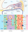
Figure 5. Schematic model of cochlear potassium circulation and the formation of the endocochlear potential. (a) K+ ions that escape from the hair cells are taken up by Deiters cells. K+ is subsequently transported via the epithelial gap junction network to the type II and type IV fibrocytes of the spiral ligament. The epithelial gap junction network consists of supporting cells, epithelial cells and the outer sulcus cells. K+ is then taken up by the type II and type IV fibrocytes and transported to the stria vascularis via the connective tissue gap junction network. The connective tissue gap junction network consists of fibrocytes, basal cells and intermediate cells. K+ is eventually released via the stria vascularis into the endolymph of the scala media. The diagram also shows the K+ concentration ([K+]) and the potential of the various cochlear fluids. (b) The figure shows the ion transport system of the stria vascularis and the spiral ligament, which are the crucial components for cochlear potassium circulation and the formation of the endocochlear potential. As in (a), the K+ concentration ([K+]) and the potential of the various cochlear fluids is shown. Abbreviations: NKCC1 (Na+ K+ 2Cl- cotransporter), TJ (tight junctions) (Figure modified after Hibino & Kurachi [53]).
Figure 6
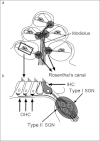
Figure 6. Schematic representation of the cochlea and the afferent innervation of the hair cells. (a) The axis of the cochlea (modiolus) harbors the spiral ganglion cells. The cell bodies are located in Rosenthal’s canal. (b) The afferent innervation of the hair cells takes place via the nerve fibres of the spiral ganglion cell neurons (SGN). Type I spiral ganglion neurons are myelinated and lead to the inner hair cells (IHC).Type II spiral ganglion neurons are not myelinated and lead to the outer hair cells (OHC). Each type II cell forms synaptic contacts with numerous outer hair cells. Type I cells, however, typically show contact with only one inner hair cell (Figure modified after Rusznák & Szücs [119], by kind permission of Springer Science + Business Media).
Figure 7
Figure 7. The ribbon synapse of the inner hair cell. The afferent synapse of the inner hair cells (HC) consists of a presynaptic active zone with the synaptic ribbon (diameter 0.2 µm), a protein nanomachine to which the synaptic vesicles (diameter 35-40 nm) are bound. The postsynaptic nerve fibre ending contains numerous ionotropic AMPA-type glutamate receptors (AMPA-R). Each active zone contains some 50 CaV1.3 calcium channels and 30 glutamate-containing synaptic vesicles (SV). Abbreviations: SC (supporting cells), (Figure modified after Fuchs et al. [120]).
Figure 8
Figure 8. Schematic representation of the efferent innervation of the organ of Corti. Myelinated mediale olivocochlear fibres (MOC) form synaptic contacts with outer hair cells (OHC). Non-myelinated lateral olivocochlear fibres (LOC) form synaptic contacts with afferent type I spiral ganglion neurons (type I SGN), which lead to the inner hair cells (IHC). Key to arrows: right-facing = efferent, left-facing = afferent, (Figure modified after Guinan [109]).

Similar articles

Cited by

References

    1. Santi PA, Tsuprun VL. Cochlear microanatomy and ultrastructure. In: Jahn AF, Santos-Sacchi J, editors. Physiology of the Ear. San Diego, CA: Singular Publishing; 2001.
    1. Slepecky NB. Cochlear structure. In: Dallos P, Popper AN, Fay R, editors. The Cochlea. New York: Springer; 1996. pp. 44–129. Available from: http://dx.doi.org/10.1007/978-1-4612-0757-3_2. - DOI - DOI
    1. Boenninghaus, Lenarz . Innenohr (Labyrinth) In: Boenninghaus, Lenarz, editors. HNO. Springer Medizin Verlag; 2007. pp. 15–19.
    1. Starlinger V, Masaki K, Heller S. Auditory physiology: Inner ear. In: Gulya AJ, Minor LB, Poe DS, editors. Glasscock-Shambaugh's Surgery of the Ear. 6th ed. People's Medical Publishing House; 2010. pp. 73–83.
    1. Rhys Evans PH, Comis SD, Osborne MP, Pickles JO, Jeffries DJ. Cross-links between stereocilia in the human organ of Corti. J Laryngol Otol. 1985;99:11–19. doi: 10.1017/S0022215100096237. Available from: http://dx.doi.org/10.1017/S0022215100096237. - DOI - DOI - PubMed