. 2012;7(4):e34140.
doi: 10.1371/journal.pone.0034140. Epub 2012 Apr 27.

Neoplastic transformation of T lymphocytes through transgenic expression of a virus host modification protein

Affiliations

Neoplastic transformation of T lymphocytes through transgenic expression of a virus host modification protein

Sílvia Cristina Paiva Almeida et al. PLoS One. 2012.

Abstract

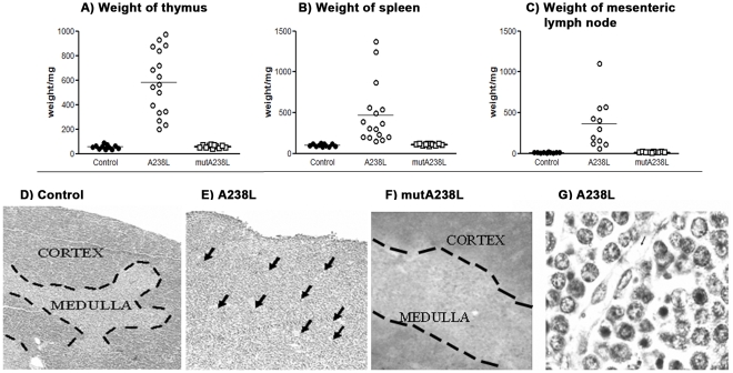
Virus host evasion genes are ready-made tools for gene manipulation and therapy. In this work we have assessed the impact in vivo of the evasion gene A238L of the African Swine Fever Virus, a gene which inhibits transcription mediated by both NF-κB and NFAT. The A238L gene has been selectively expressed in mouse T lymphocytes using tissue specific promoter, enhancer and locus control region sequences for CD2. The resulting two independently derived transgenic mice expressed the transgene and developed a metastasic, angiogenic and transplantable CD4(+)CD8(+)CD69(-) lymphoma. The CD4(+)CD8(+)CD69(-) cells also grew vigorously in vitro. The absence of CD69 from the tumour cells suggests that they were derived from T cells at a stage prior to positive selection. In contrast, transgenic mice similarly expressing a mutant A238L, solely inhibiting transcription mediated by NF-κB, were indistinguishable from wild type mice. Expression of Rag1, Rag2, TCRβ-V8.2, CD25, FoxP3, Bcl3, Bcl2 l14, Myc, IL-2, NFAT1 and Itk, by purified CD4(+)CD8(+)CD69(-) thymocytes from A238L transgenic mice was consistent with the phenotype. Similarly evaluated expression profiles of CD4(+)CD8(+) CD69(-) thymocytes from the mutant A238L transgenic mice were comparable to those of wild type mice. These features, together with the demonstration of (mono-)oligoclonality, suggest a transgene-NFAT-dependent transformation yielding a lymphoma with a phenotype reminiscent of some acute lymphoblastic lymphomas.


Conflict of interest statement

Competing Interests: The authors have declared that no competing interests exist.

Figures

Figure 1
Figure 1. Expression of the A238L transgene confirmed by RT-PCR and by detection of the protein.
A) B) RT-PCR for the A238L gene is negative in transgenic splenic B cells after MACS purification (lane 1), and positive in total splenocytes (lane 2) and total thymocytes (lane 3). The upper bands are the A238L PCR product, and the bottom bands are the tubulin controls. C) A similar RT-PCR analysis of two cell lines independently established from the thymuses of two different A238L T cell transgenic mice (lanes 1 and 2), and of a tumour that developed as the result of subcutaneous injection of thymic transgenic cells (lane 3). The upper bands are the A238L PCR product, and the bottom bands are the tubulin controls. Immunofluorescent staining of control (D), A238L (E) and mutA238L (F) T cell restricted transgenic thymus cryostat sections using a rat monoclonal antibody against the HA immunotag upstream the A238L transgene, followed by a goat anti-rat FITC (magnification 40X). The results are representative of at least 5 mice per founder and per genotype.
Figure 2
Figure 2. Enlargement and loss of organization of lymphoid organs in A238L T cell transgenic mice.
Weight of the Thymus (A), Spleen (B) and Mesenteric Lymph node (C) of control, A238L and mutA238L T cell restricted transgenic mice. Each symbol represents an animal and black and white circles and white squares correspond to 11 control, 17 A238L and 11 mutA238L transgenic mice, respectively. Haematoxylin eosin stained sections of control mouse thymus (D) showing the typical cortex, with densely packed cells clearly differentiated from the medulla with less densely packed cells, and transgenic A238L thymus (E), containing blood vessels (see arrows) and lacking structural organization. The infiltrate of the thymus is accompanied by an increase in vascularisation (see arrows) (magnification 10X). At higher magnification, the transgenic thymus was seen to consist of typical lymphoblasts and significantly more dense apoptotic nuclei than the control thymus (G) (Magnification 40X). Vascularisation of the T cell restricted A238L transgenic thymus is also shown in the photograph (G), (note: blood vessel with its endothelial cell wall and shadows of red cells in the lumen). The results are representative of at least 5 mice per founder and per genotype.
Figure 3
Figure 3. Phenotype of thymocytes from T cell restricted A238L transgenic mice.
A) Surface phenotype of thymic cell suspensions of control littermates (left), Tg A238L (middle) and Tg mut A238L mice (right). Cells were stained with antibodies specific for CD4-PE, CD8-APC and CD69-FITC cell surface markers. Results are presented as CD4 versus CD8 (A) and the indicated CD4+CD8+ cells were then examined for CD69 expression (B, presented as CD4 versus CD69, and graphs E and F). The same cells were also stained with CD4-PE, CD8-APC, CD25-CyChr and CD44-FITC, and the expression of CD25 and CD44 on CD4CD8 double positive cells was determined (C, presented as CD25 versus CD44). The analysis was also performed for splenic and lymph node cell suspensions and the same results were obtained (not shown). The reduced expression of CD3-APC and TCRαβ-Alexa488 on T cell restricted A238L transgenic mouse thymocytes was demonstrated by staining with antibodies specific for CD3 and TCRαβ (D, presented as CD3 versus TCRαβ, graphs E and F). The results are for groups of 5 mice per genotype and are representative of at least three independent experiments.
Figure 4
Figure 4. Demonstration of endothelial cells and VEGF synthesis in the thymus of the T cell restricted A238L transgenic mouse.
Cryostat sections of thymus of control littermates (left column), A238L Tg (middle column) and mutA238L Tg (right column) mice were stained with antibodies specific for VEGF (upper row) (magnification 20X) and endothelial cells (CD31) (lower row) (magnification 40X). The results are representative of at least 5 mice per founder and per genotype.
Figure 5
Figure 5. Southern-blot analysis of TCRβ clonality of the thymic transgenic cells in the A238L T cell restricted transgenic mice.
30 µg of DNA from the thymus of transgenic mice (lanes 1 to 6), of transgenic thymic in vitro established cell lines (lanes 7 and 8) and of wild type control mice (lane 9) was digested with enzymes A) HindIII, B) PvuII, C) EcoRV and D) HpaI, as indicated on the top of the gels. Lanes 1 to 4 and 5 to 6 are of progeny mice of Founders 1 and 2, respectively. The in vitro established cell lines, corresponding to lanes 7 and 8 were derived from Founders 2 and 1, respectively, and do not correspond to the mouse thymus DNA samples analyzed on the gel. Lane MW indicates the molecular weight marker used and the corresponding values are indicated in the text box on its left.
Figure 6
Figure 6. Metastasis of thymocytes from T cell restricted A238L transgenic thymus.
The figure shows sections of lymphoid organs stained with Hematoxylin/Eosin (magnification 10 X). There is an exuberant infiltration ofmesenteric lymph nodes, with a corresponding loss of tissue organization. (Not shown: There is also an infiltration of other tissues in areas around arteries and blood vessels, such as kidney, liver and lung and morphology of the infiltrating lymphocytes is similar to that observed in the transgenic thymuses) The results are representative of at least 5 mice per founder and per genotype.
Figure 7
Figure 7. Expression analysis of CD4+CD8+CD69 thymocytes demonstrates selective effect of the expression of the A238L transgene in the A238L transgenic mice.
Semi-quantitative PCR analysis of the cDNA prepared from the mRNA of the purified CD4+CD8+CD69 thymic cells of A238L and mutA238L transgenic and control mice shows that the A238L transgenic mice display a distinctive pattern of expression for a group of selective genes which correlates to the phenotype of the mice. β-Globin was used as the housekeeping gene. The results presented correspond to a pool of 5 mice per genotype.

Similar articles

Cited by

References

    1. Finlay BB, McFadden G. 2006. (2006) Anti-immunology: evasion of the host immune system by bacterial and viral pathogens. Cell 124: 767–782. Available: http://www.ncbi.nlm.nih.gov/pubmed/16497587.
    1. Vischer HF, Vink C, Smit MJ. 2006. (2006) A viral conspiracy: hijacking the chemokine system through virally encoded pirated chemokine receptors. Current topics in microbiology and immunology 303: 121–154. Available: http://www.ncbi.nlm.nih.gov/pubmed/16570859. - PubMed
    1. Loo YM, Gale M. 2007. (2007) Viral regulation and evasion of the host response. Current topics in microbiology and immunology 316: 295–313. Available: http://www.ncbi.nlm.nih.gov/pubmed/17969453.
    1. Unterholzner L, Bowie AG. 2008. (2008) The interplay between viruses and innate immune signaling: recent insights and therapeutic opportunities. Biochemical pharmacology 75: 589–602. Available: http://www.ncbi.nlm.nih.gov/pubmed/17868652. - PubMed
    1. Bonjardim CA, Ferreira PCP, Kroon EG. 2009. (2009) Interferons: signaling, antiviral and viral evasion. Immunology letters 122: 1–11. Available: http://www.ncbi.nlm.nih.gov/pubmed/19059436.

Publication types

MeSH terms