. 2012;7(4):e34303.
doi: 10.1371/journal.pone.0034303. Epub 2012 Apr 27.

JKA97, a novel benzylidene analog of harmine, exerts anti-cancer effects by inducing G1 arrest, apoptosis, and p53-independent up-regulation of p21

Affiliations

JKA97, a novel benzylidene analog of harmine, exerts anti-cancer effects by inducing G1 arrest, apoptosis, and p53-independent up-regulation of p21

Xinyi Yang et al. PLoS One. 2012.

Abstract

JKA97, a benzylidene analog of harmine, has been found to be a promising drug candidate for human cancer therapy, although the underlying molecular mechanisms have not been fully demonstrated. In this study, we evaluated the effects of JKA97 on human breast cancer cells in vitro and in vivo. JKA97 inhibited the growth and proliferation of MCF7 (p53 wild-type), MCF7 (p53 knockdown), and MDA-MB-468 (p53 mutant) cells in a dose-dependent manner. Treatment with JKA97 arrested breast cancer cells in G1 phase and induced apoptosis. JKA97 also significantly suppressed the growth of MCF7 and MDA-MB-468 xenograft tumors. It regulated the expression levels of G1 phase regulators, such as p21, p27, cyclinE, and cylinD1. JKA97 activated p21 transcription, independent of p53, but had little effect on p21 protein stability/degradation. In summary, our results suggest that JKA97 inhibits human breast cancer cell growth through activating p21, independent of p53, which provides a basis for developing this compound as a novel drug for human breast cancer therapy.


Conflict of interest statement

Competing Interests: The authors have declared that no competing interests exist.

Figures

Figure 1
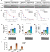
Figure 1. In vitro anticancer activities of JKA97 against breast cancer cells.
(A) Chemical structure of JKA97. (B) Concentrations of JKA97 inducing 50% growth inhibition (IC50) in breast cancer cells, relative to the corresponding controls, based on the MTT assay. MCF7, MDA-MB-468, and MCF7 p53KD cells were exposed to various concentrations of JKA97 for 72 hrs. (C) Anti-proliferative effects of JKA97 on breast cancer cells. Cells were exposed to various concentrations of JKA97 for 48 hrs, followed by the BrdUrd incorporation assay. The proliferation index was calculated against untreated control cells (*P<0.05). (D) Induction of apoptosis in breast cancer cells by JKA97. Cells were exposed to various concentrations of JKA97 for 24 hrs, followed by measurement of apoptosis by Annexin V assay. The apoptotic index was calculated against untreated control cells (*P<0.05). (E) Effects of JKA97 on the cell cycle distribution of breast cancer cells. Cells were exposed to various concentrations of JKA97 for 24 hrs, followed by measurement DNA contents by flow cytometry. The cell cycle distribution was evaluated by comparing with that of control cells (*P<0.05). All the assays were performed in triplicate. Results were from at least three separate, repeated experiments.
Figure 2
Figure 2. In vivo anticancer activity of JKA97 against breast cancer cells.
JKA97 was administered by i.p. injection to nude mice bearing MCF7 (A1) or MDA-MB-468 (B1) xenograft tumors. For mice bearing MCF7 xenograft tumor, treatment groups received JKA97 at doses of 5 mg/kg/day or 25 mg/kg/day, 5 days/week, for 6 weeks; for mice bearing MDA-MB-468 xenograft tumor, treatment groups received doses at 5 mg/kg/day or 10 mg/kg/day, 5 days/week, for 18 days. Control groups received vehicle only. Animals were also monitored for changes in body weight as a surrogate marker for toxicity when it was administered to nude mice bearing (A2) MCF7 or (B2) MDA-MB-468 xenograft tumors. At the end of the experiments, xenograft tumors were removed and taken a photograph (A3 and B3).
Figure 3
Figure 3. Effects of JKA97 on the expression of cell cycle related proteins.
(A) MCF7, MDA-MB-468, and MCF7 p53KD cells were exposed to various concentrations of JKA97 for 24 hrs, and the expression of p53 and p21 proteins was determined by Western blot analysis. (B) Cells were exposed to 10 µM of JKA97 for the indicated time, and the expression of p53 and p21 proteins was determined by Western blot analysis. β-actin was used as an equal-loading control of samples.
Figure 4
Figure 4. Effects of JKA97 on p21 expression.
(A1) MCF7, MDA-MB-468, and MCF7 p53KD cells were exposed to various concentrations of JKA97 or vehicle for 24 hrs, followed by exposure to protein synthesis inhibitor cycloheximide (CHX, 10 µg/mL). p21 protein expression was detected by Western blotting at different times after exposure of CHX. (A2) The graph shows the quantification of the Western blotting data. MCF7 (B1), MDA-MB-468 (B2) and MCF7 p53KD (B3) Cells were exposed to various concentrations of JKA97 or vehicle for 24 hrs, and total RNA were extracted followed by reverse transcription, and detection mRNA level of p21 by Real-time quantification PCR and Quantification RT-PCR, normalized by mRNA level of GAPDH. (C) Cells were transfected with p21 promoter luciferase reporter plasmid and a Renilla luciferase reporter together for 12 hrs, followed by treatment of 10 µM JKA97 or vehicle for an additional 24 hrs. The reporter activity was normalized to the corresponding Renilla luciferase reporter. The luciferase assay was performed in triplicate. Statistical significance was determined compared with control (*P<0.05).

Similar articles

Cited by

References

    1. Jemal A, Bray F, Center MM, Ferlay J, Ward E, et al. Global cancer statistics. CA Cancer J Clin. 2011;61:69–90. - PubMed
    1. Nathanson KL, Domchek SM. Therapeutic approaches for women predisposed to breast cancer. Annu Rev Med. 2011;62:295–306. - PubMed
    1. Oakman C, Santarpia L, Di Leo A. Breast cancer assessment tools and optimizing adjuvant therapy. Nat Rev Clin Oncol. 2010;7:725–732. - PubMed
    1. Eskelinen M, Hippelainen M, Kettunen J, Salmela E, Penttila I, et al. Clinical value of serum tumour markers TPA, TPS, TAG 12, CA 15-3 and MCA in breast cancer diagnosis; results from a prospective study. Anticancer Res. 1994;14:699–703. - PubMed
    1. Koehn FE, Carter GT. The evolving role of natural products in drug discovery. Nat Rev Drug Discov. 2005;4:206–220. - PubMed

Publication types

MeSH terms