. 2012;7(4):e34962.
doi: 10.1371/journal.pone.0034962. Epub 2012 Apr 25.

Ss-Sl2, a novel cell wall protein with PAN modules, is essential for sclerotial development and cellular integrity of Sclerotinia sclerotiorum

Affiliations

Ss-Sl2, a novel cell wall protein with PAN modules, is essential for sclerotial development and cellular integrity of Sclerotinia sclerotiorum

Yang Yu et al. PLoS One. 2012.

Abstract

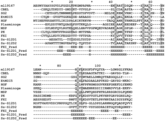
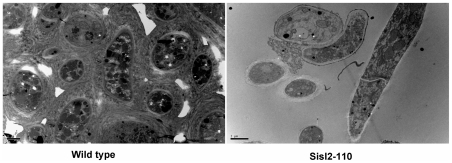
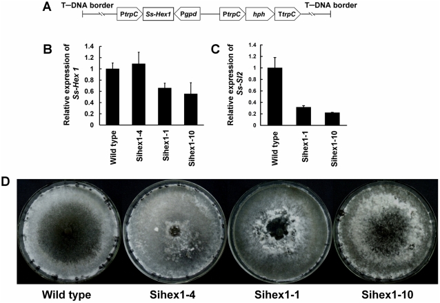
The sclerotium is an important dormant body for many plant fungal pathogens. Here, we reported that a protein, named Ss-Sl2, is involved in sclerotial development of Sclerotinia sclerotiorum. Ss-Sl2 does not show significant homology with any protein of known function. Ss-Sl2 contains two putative PAN modules which were found in other proteins with diverse adhesion functions. Ss-Sl2 is a secreted protein, during the initial stage of sclerotial development, copious amounts of Ss-Sl2 are secreted and accumulated on the cell walls. The ability to maintain the cellular integrity of RNAi-mediated Ss-Sl2 silenced strains was reduced, but the hyphal growth and virulence of Ss-Sl2 silenced strains were not significantly different from the wild strain. Ss-Sl2 silenced strains could form interwoven hyphal masses at the initial stage of sclerotial development, but the interwoven hyphae could not consolidate and melanize. Hyphae in these interwoven bodies were thin-walled, and arranged loosely. Co-immunoprecipitation and yeast two-hybrid experiments showed that glyceraldehyde-3-phosphate dehydrogenase (GAPDH), Woronin body major protein (Hex1) and elongation factor 1-alpha interact with Ss-Sl2. GAPDH-knockdown strains showed a similar phenotype in sclerotial development as Ss-Sl2 silenced strains. Hex1-knockdown strains showed similar impairment in maintenance of hyphal integrity as Ss-Sl2 silenced strains. The results suggested that Ss-Sl2 functions in both sclerotial development and cellular integrity of S. sclerotiorum.


Conflict of interest statement

Competing Interests: The authors have declared that no competing interests exist.

Figures

Figure 1
Figure 1. Multiple alignment of sequences of Ss-Sl2D1, Ss-Sl2D2 and representative PAN modules using ClustalX.
The selected PAN modules are from a Mesorhizobium loti mll9167 protein (mll9167, BAB54559, residues 117–194), Phytophthora parasitica CBEL protein (CBEL, CAA65843, residues 197–268), Ipomoea trifida secreted glycoprotein 2 (SG2, AAA97902, residues 348–425), Eimeria tenella microneme protein 5 (EtMIC5, CAB52368, residues 713–781), human macrophage stimulating protein (MSP, BAH12774, residues 21–105), human plasminogen (Plasminogen, AAA36451, residues 20–98), human hepatocyte growth factor (HGF, AAA52648, residues 37–123), human prekallikrein (PK, AAY40900, residues 21–103), and human coagulation factor XI (FXI, AAA51985, residues 20–103). Conserved amino acids are shown with a shaded background. The secondary structural elements (H for alpha helix, E for beta strand) from the PAN/apple domains of FXI (FXI_Pred), Ss-Sl2D1 (Ss-Sl2D1_Pred) and Ss-Sl2D2 (Ss-Sl2D1_Pred) predicted with the PSIPRED program are the last three sequences in the multiple alignments.
Figure 2
Figure 2. Subcellular location of Ss-Sl2 in S. sclerotiorum.
Immunogold labeling of ultrathin sections from (A) hyphae at the early stage of vegetative growth (cultured on PDA plates for 1 day), (B) hyphae at the later stage of vegetative growth (cultured for 3 days), (C) sclerotial initial (cultured for 4 days), (D) sclerotia just starting to accumulate melanin and consolidate (cultured for 6 days), and (E) mature sclerotia (cultured for 7 days) with anti-Ss-Sl2 polyclonal antibodies, or from hyphae with preimmune serum (F) are shown. The gold particles are visible on the cell walls and septa in a patchy distribution. W, cell wall; S, septa; WB, Woronin body. Bar = 1 μm.
Figure 3
Figure 3. Ss-Sl2 was detected in cytoplasm and cell wall of S. sclerotiorum.
The cell wall protein and cytoplasm protein in mycelium were extracted respectively (50 μg) and subjected to western blot analysis with anti-Ss-Sl2 polyclonal antibodies.
Figure 4
Figure 4. Real-time RT-PCR analysis of Ss-Sl2 transcript in different sclerotial developmental stages of S. sclerotiorum.
S0 (early)  =  the early stage of vegetative growth (cultured on PDA plates for 1 day); S0 (later)  =  the later stage of vegetative growth (cultured for 3 days); S1 =  the initiation stage of sclerotial development (cultured for 4 days); S2 =  condensation stage (cultured for 5 days); S4 =  consolidation stage (cultured for 6 days); S6 =  maturation stage (cultured for 7 days). The expression level of Ss-Sl2 cDNA measured by RT-PCR was normalized to that of actin cDNA in extracts from each developmental stage. The abundance of cDNA from S0 (early) samples was assigned a value of 1. Bars indicate standard error.
Figure 5
Figure 5. Construction of an Ss-Sl2 RNAi vector (pSisl2) and functional analysis of Ss-Sl2 silenced strains.
(A) A 310 bp fragment of Ss-Sl2 was inserted in the sense orientation between an Aspergillus nidulans trpC promoter and an intron from Gibberella zeae, and the same fragment of Ss-Sl2 was inserted in antisense orientations between this intron and the trpC terminator. (B) Expression level of Ss-Sl2 in isolates containing pSisl2 and in the wild type strain were determined by real-time RT-PCR. The expression level of Ss-Sl2 cDNA was normalized to that of actin cDNA in extracts from each strain. The abundance of cDNA from the wild type was assigned a value of 1. Bars indicate standard error. (C) Phenotype of the wild type strain and Ss-Sl2 gene-silenced transformants.
Figure 6
Figure 6. Ultrastructural analysis of sclerotia produced by the wild type and Sisl2-110.
The inner structure of sclerotia produced by the wild type showing a compact structure and by Sisl2-110 showing a loose structure, both observed by TEM. Bar = 2 μm.
Figure 7
Figure 7. Expression level of a melanin biosynthesis associated polyketide synthase-encoding gene (Ss-Pks1) in Ss-Sl2 gene silenced strains of S. sclerotiorum by real-time RT-PCR.
The expression level of Ss-Pks1 cDNA was normalized to that of actin cDNA in extracts from each strain. The abundance of cDNA from the wild type was assigned a value of 1. Bars indicate standard error.
Figure 8
Figure 8. Growth characteristics and analysis of Ss-Sl2 silenced strains of S. sclerotiorum.
(A) The inhibition of hyperosmotic stress and sorbose to the hyphal growth rate of Ss-Sl2 silenced strains and the wild type. *Significantly different from the wild type strain (P<0.05). (B) Cytoplasmic bleeding at the hyphal tips of Sisl2-110 when cultured on medium with 5% sorbose for 2 d. Bar = 50 μm.
Figure 9
Figure 9. Interaction of Ss-Sl2 and GAPDH, EF-1α and Hex1 in yeast Y2HGold cells.
The Ss-Sl2 region code for amino acid 17–352 (without signal peptide) was inserted into pGBKT7 to get the bait plasmid. The cDNA of GAPDH, EF-1α and Hex1 were inserted into pGADT7 to obtain the prey plasmids. The prey plasmids respectively were co-transformed into Y2HGold with the bait plasmid. Transformed Y2HGold cells grown on the SD/-Leu-Trp, SD/-Leu-Trp-His-Ade and SD/-Leu-Trp-His-Ade with X-α-gal. pGBKT7 and pGADT7 are bait and prey vectors without inserts. pGBKT7-53 and pGADT7-T encode two fusion proteins that are known to interact (Clontech).
Figure 10
Figure 10. Construction of Ss-Gpd RNAi vector (pSigapdh) and analysis of Ss-Gpd silenced strains.
(A) A 538-bp fragment of Ss-Gpd was inserted between the N. crassa trpC promoter PtrpC and A. nidulans gpd promoter Pgpd. (B) Expression level of Ss-Gpd in isolates containing the pSigapdh and in the wild type determined by real-time RT-PCR. The expression level of Ss-Gpd cDNA was normalized to that of actin cDNA in extracts from each strain. The abundance of cDNA from the wild type was assigned a value of 1. Bars indicate standard error. (C) Expression level of Ss-Sl2 in Ss-Gpd silenced strains and in the wild type determined by real-time RT-PCR. The expression level of Ss-Sl2 cDNA was normalized to that of actin cDNA in extracts from each strain. The abundance of cDNA from the wild type was assigned a value of 1. Bars indicate standard error. (D) Phenotype of the wild type and Ss-Gpd gene-silenced transformants.
Figure 11
Figure 11. Functional analysis of Ss-Hex1 with RNAi technology.
(A) A 465-bp fragment of Ss-Hex1 was inserted between the N. crassa trpC promoter PtrpC and A. nidulans gpd promoter Pgpd. (B) Expression level of Ss-Hex1 in strains containing the pSihex1 and wild type strain using the real-time RT-PCR. The expression level of Ss-Hex1 cDNA was normalized to that of actin cDNA in extracts from each isolate. The abundance of cDNA from the wild type was assigned a value of 1. Bars indicate standard error. (C) Expression level of Ss-Sl2 in Ss-Hex1 silenced strains and in the wild type determined by real-time RT-PCR. The expression level of Ss-Sl2 cDNA was normalized to that of actin cDNA in extracts from each strain. The abundance of cDNA from the wild type was assigned a value of 1. Bars indicate standard error. (D) Phenotype of the wild type and Ss-Hex1 gene-silenced transformants.
Figure 12
Figure 12. Growth characteristics and analysis of Ss-Hex1 silenced strains.
(A) The inhibition of hyperosmotic stress and sorbose to the hyphal growth rate of Ss-Hex1 silenced strains and the wild type. *Significantly different from the wild type strain (P<0.05). (B) Microscopic observation of hyphal tips and forming branches for Sihex1-10 and the wild type on PDA and PDA with 5% sorbose. Bar = 100 μm.

Similar articles

Cited by

References

    1. Boland GJ, Hall R. Index of plant hosts of Sclerotinia sclerotiorum. Can J Plant Pathol. 1994;16:93–100.
    1. Bolton M, Thomma BPHJ, Nelson B. Sclerotinia sclerotiorum (Lib.) de Bary: biology and molecular traits of a cosmopolitan pathogen. Mol Plant Pathol. 2006;7:1–16. - PubMed
    1. Steadman JR. Control of plant diseases caused by Sclerotinia species. Phytopathology. 1979;69:904–907.
    1. Roper M, Seminara A, Bandi MM, Cobb A, Dillard HR, et al. Dispersal of fungal spores on a cooperatively generated wind. Proc Natl Acad Sci U S A. 2010;107:17474–17479. - PMC - PubMed
    1. Kosasih BD, Willetts HJ. Ontogenetic and histochemical studies of the apothecium of Sclerotinia sclerotiorum. Ann Bot. 1975;39:185–191.

Publication types

MeSH terms