

Hyperlipidemia and atherosclerotic lesion development in Ldlr-deficient mice on a long-term high-fat diet
- PMID: 22558236
- PMCID: PMC3338468
- DOI: 10.1371/journal.pone.0035835
Hyperlipidemia and atherosclerotic lesion development in Ldlr-deficient mice on a long-term high-fat diet
Abstract
Background: Mice deficient in the LDL receptor (Ldlr(-/-) mice) have been widely used as a model to mimic human atherosclerosis. However, the time-course of atherosclerotic lesion development and distribution of lesions at specific time-points are yet to be established. The current study sought to determine the progression and distribution of lesions in Ldlr(-/-) mice.
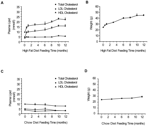
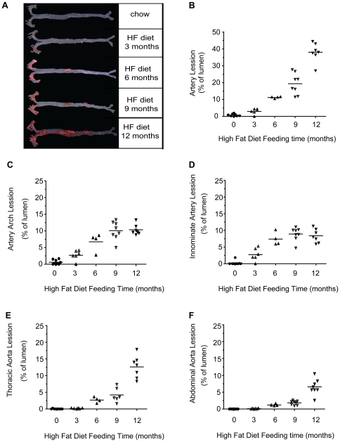
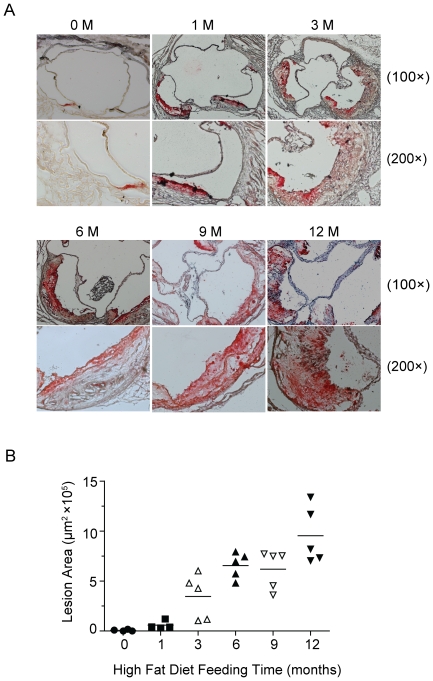
Methodology/principal findings: Ldlr-deficient mice fed regular chow or a high-fat (HF) diet for 0.5 to 12 months were analyzed for atherosclerotic lesions with en face and cross-sectional imaging. Mice displayed significant individual differences in lesion development when fed a chow diet, whereas those on a HF diet developed lesions in a time-dependent and site-selective manner. Specifically, mice subjected to the HF diet showed slight atherosclerotic lesions distributed exclusively in the aortic roots or innominate artery before 3 months. Lesions extended to the thoracic aorta at 6 months and abdominal aorta at 9 months. Cross-sectional analysis revealed the presence of advanced lesions in the aortic sinus after 3 months in the group on the HF diet and in the innominate artery at 6 to 9 months. The HF diet additionally resulted in increased total cholesterol, LDL, glucose, and HBA1c levels, along with the complication of obesity.
Conclusions/significance: Ldlr-deficient mice on the HF diet tend to develop site-selective and size-specific atherosclerotic lesions over time. The current study should provide information on diet induction or drug intervention times and facilitate estimation of the appropriate locations of atherosclerotic lesions in Ldlr(-/-) mice.
Conflict of interest statement
Figures

References
-
- Glass CK, Witztum JL. Atherosclerosis. the road ahead. Cell. 2001;104:503–516. - PubMed
-
- Rader DJ, Daugherty A. Translating molecular discoveries into new therapies for atherosclerosis. Nature. 2008;451:904–913. - PubMed
-
- Zadelaar S, Kleemann R, Verschuren L, de Vries-Van der Weij J, van der Hoorn J, et al. Mouse models for atherosclerosis and pharmaceutical modifiers. Arterioscler Thromb Vasc Biol. 2007;27:1706–1721. - PubMed
-
- Nakashima Y, Plump AS, Raines EW, Breslow JL, Ross R. ApoE-deficient mice develop lesions of all phases of atherosclerosis throughout the arterial tree. Arterioscler Thromb. 1994;14:133–140. - PubMed
-
- Reddick RL, Zhang SH, Maeda N. Atherosclerosis in mice lacking apo E. Evaluation of lesional development and progression. Arterioscler Thromb. 1994;14:141–147. - PubMed
Publication types
MeSH terms
Substances
LinkOut - more resources
Full Text Sources
Other Literature Sources
Medical
Molecular Biology Databases
Research Materials
Miscellaneous
