. 2012;7(4):e35882.
doi: 10.1371/journal.pone.0035882. Epub 2012 Apr 27.

Deletion of Dicer in smooth muscle affects voiding pattern and reduces detrusor contractility and neuroeffector transmission

Affiliations

Deletion of Dicer in smooth muscle affects voiding pattern and reduces detrusor contractility and neuroeffector transmission

Mardjaneh Karbalaei Sadegh et al. PLoS One. 2012.

Abstract

MicroRNAs have emerged as important regulators of smooth muscle phenotype and may play important roles in pathogenesis of various smooth muscle related disease states. The aim of this study was to investigate the role of miRNAs for urinary bladder function. We used an inducible and smooth muscle specific Dicer knockout (KO) mouse which resulted in significantly reduced levels of miRNAs, including miR-145, miR-143, miR-22, miR125b-5p and miR-27a, from detrusor preparations without mucosa. Deletion of Dicer resulted in a disturbed micturition pattern in vivo and reduced depolarization-induced pressure development in the isolated detrusor. Furthermore, electrical field stimulation revealed a decreased cholinergic but maintained purinergic component of neurogenic activation in Dicer KO bladder strips. The ultrastructure of detrusor smooth muscle cells was well maintained, and the density of nerve terminals was similar. Western blotting demonstrated reduced contents of calponin and desmin. Smooth muscle α-actin, SM22α and myocardin were unchanged. Activation of strips with exogenous agonists showed that depolarization-induced contraction was preferentially reduced; ATP- and calyculin A-induced contractions were unchanged. Quantitative real time PCR and western blotting demonstrated reduced expression of Cav1.2 (Cacna1c). It is concluded that smooth muscle miRNAs play an important role for detrusor contractility and voiding pattern of unrestrained mice. This is mediated in part via effects on expression of smooth muscle differentiation markers and L-type Ca(2+) channels in the detrusor.


Conflict of interest statement

Competing Interests: The authors have declared that no competing interests exist.

Figures

Figure 1
Figure 1. Reduced levels of detrusor miRNAs and maintained wet weight in Dicer KO urinary bladder.
Highly expressed miRNAs were selected in an array experiment and analyzed here by qPCR in control (black bars) and Dicer KO (white bars) urinary bladders excised 10 weeks following tamoxifen treatment (A) (n = 6). MiR-451 is generated in Dicer independent manner and was included as a negative control. The bladder wet weights, bladder to body weight ratios, and body weights of control and Dicer KO bladders is shown in B-D (n = 13).
Figure 2
Figure 2. Disturbed micturition pattern and impaired pressure generation in the isolated bladder.
Voided urine was collected on filter papers. Panel A shows filter papers photographed under UV light. Panel B shows summarized data. Panel C shows pressure of isolated and cannulated bladders during 5 min of stimulation with 60 mM K+ at different volumes (6–8).
Figure 3
Figure 3. Reduced active stress in Dicer KO detrusor strips.
Active (A) and passive (B) length-tension relationships were generated using strips from control and Dicer KO bladders. The muscle strips were stimulated in 60 mM K+ and relaxed in nominally calcium-free solution. Stress (C) was calculated using the length and weight of the individual preparations. The calculated circumference at which the bladder generated maximal active force (L0) was not different in Dicer KO bladders (D). Stress at L0 is shown in E (n = 11).
Figure 4
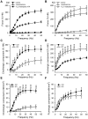
Figure 4. Deletion of Dicer in smooth muscle impairs the cholinergic component of the neurogenic contraction.
Detrusor muscle strips from control and Dicer KO mice were activated by electrical field stimulation in the absence and presence of scopolamine and after desensitization of purinergic receptors using α,β-methylene-ATP in the continued presence of scopolamine. Full frequency response curves in control conditions, in the presence of scopolamine (1 µM), and after desensitization of purinergic receptors using α,β-methylene-ATP (10 µM) in the continued presence of scopolamine are shown for control and Dicer KO bladders in A and B, respectively, 10 weeks following Tamoxifen treatment. The cholinergic component of activation (C) was calculated by subtracting the force in the presence of scopolamine from force in control conditions. The purinergic component of activation (D) was calculated by subtracting residual force (after α,β-methylene-ATP and in the presence of scopolamine) from force in the presence of scopolamine. The stress (force per cross-sectional area) for the different components was calculated using absolute force values, strip length, strip weight, and assuming a density of 1.06; the resulting data is shown in E-F. Stress was then calculated for the scopolamine sensitive (cholinergic, E) and the α,β-methylene-ATP sensitive (purinergic, F) components (n = 10).
Figure 5
Figure 5. Electron microscopy reveals normal cell morphology and innervation but increased distance between cells in Dicer KO urinary bladder.
Smooth muscle cells in control (A, C, E) and Dicer KO (B, D, F) bladders were analyzed by electron microscopy. 600 EM micrographs in total were used for quantitative analysis of the density of caveolae (G), the relative proportion of each cell profile that was occupied by dense bands (H), the cell cross sectional area (I), the distance between nerve terminals (J), the width of the synaptic cleft at sites with no Schwann cell coating (K), and the cell to cell distance (L) (n = 3–4).
Figure 6
Figure 6. Reduced contents of calponin and desmin in Dicer KO detrusor.
Expression of the differentiation related proteins calponin (A), desmin (B), smooth muscle α-actin (C), SM22 (D), and myocardin (E) was analyzed in control (black bars) and Dicer KO (white bars) bladders by western blotting 10 weeks post tamoxifen treatment. Original blots are shown below the individual bar graphs. HSP90 was used as loading control throughout (n = 6–8). Transcript levels for selected genes were examined in control and Dicer KO detrusor by qPCR 10 weeks post tamoxifen treatment. Primers for genes encoding calponin (Cnn1, F), Desmin (Des, G), smooth muscle α-actin (Acta2, H), SM22 (Tagln, I), Myocardin (Myocd, J) were used (n = 5–11).
Figure 7
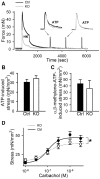
Figure 7. Selective reduction of depolarization-induced stress in Dicer KO bladder.
Panel A shows original force records of control (black line) and Dicer KO (gray line) bladder strips. Following contraction in response to 60 mM K+ (HK) and relaxation, 3 mM ATP was added every 10 min. The preparations were washed three times following each ATP challenge. Insets show representative ATP responses on an expanded time scale (n = 4–7). B and C show summarized data for the peak ATP- and α,β-methylene-ATP-induced stress in control (black bar) and Dicer KO (white bar) bladders. Panel D shows cumulative concentration-response relationship for carbachol for control and Dicer KO bladders (n = 10–11).
Figure 8
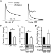
Figure 8. Reduced expression of L-type Ca2+ channels in Dicer KO bladder.
Panel A shows contractile responses to 60 mM K+ and 3 mM ATP, respectively, before (black trace) and after (gray trace) addition of the L-type Ca2+ channel blocker nifedipine (1 µM). Both K+ (not shown) and ATP (Figure 7A) responses were highly reproducible (n = 4). Panels B and C show the mRNA and protein levels for the pore-forming subunit of the L-type Ca2+ channel (Cacna1c and Cav1.2) in control (black bars) and Dicer KO (white bars) bladder. Expression of CamKIIδ is shown in D (n = 6–10).

References

    1. Andersson KE. Handb Exp Pharmacol; 2011. Muscarinic acetylcholine receptors in the urinary tract. pp. 319–344. - PubMed
    1. Fry CH, Meng E, Young JS. The physiological function of lower urinary tract smooth muscle. Auton Neurosci. 2010;154:3–13. - PubMed
    1. Gabella G. The structural relations between nerve fibres and muscle cells in the urinary bladder of the rat. J Neurocytol. 1995;24:159–187. - PubMed
    1. Ding HL, Ryder JW, Stull JT, Kamm KE. Signaling processes for initiating smooth muscle contraction upon neural stimulation. J Biol Chem. 2009;284:15541–15548. - PMC - PubMed
    1. Matsui M, Motomura D, Karasawa H, Fujikawa T, Jiang J, et al. Multiple functional defects in peripheral autonomic organs in mice lacking muscarinic acetylcholine receptor gene for the M3 subtype. Proc Natl Acad Sci U S A. 2000;97:9579–9584. - PMC - PubMed

Publication types

MeSH terms