. 2012;7(4):e35905.
doi: 10.1371/journal.pone.0035905. Epub 2012 Apr 27.

Overexpression of angiopoietin-1 increases CD133+/c-kit+ cells and reduces myocardial apoptosis in db/db mouse infarcted hearts

Affiliations

Overexpression of angiopoietin-1 increases CD133+/c-kit+ cells and reduces myocardial apoptosis in db/db mouse infarcted hearts

Heng Zeng et al. PLoS One. 2012.

Abstract

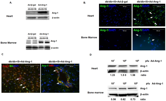
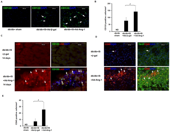
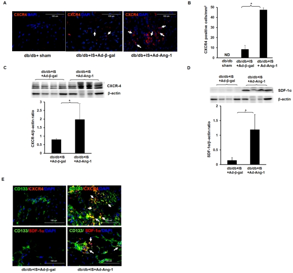
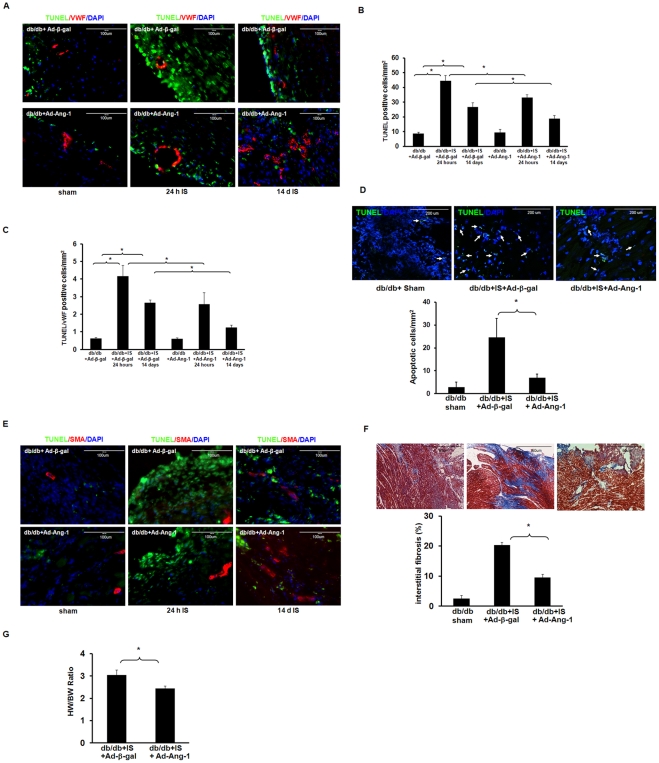
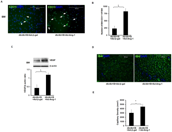
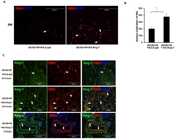
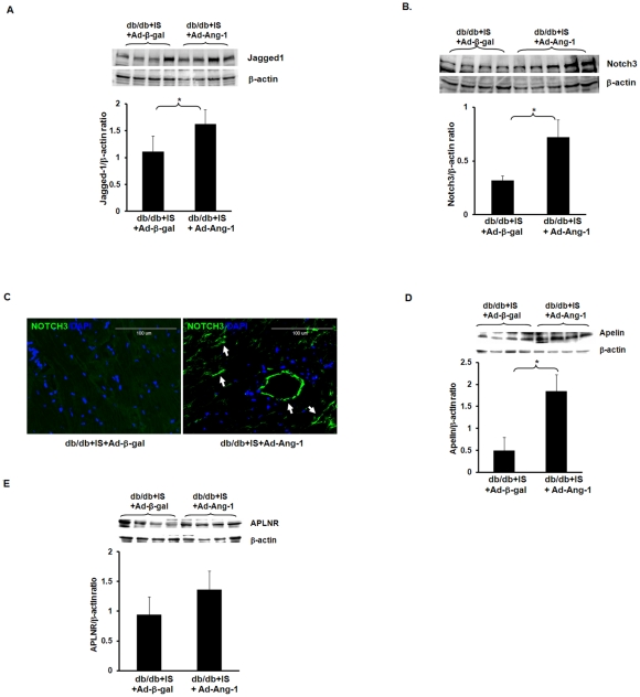
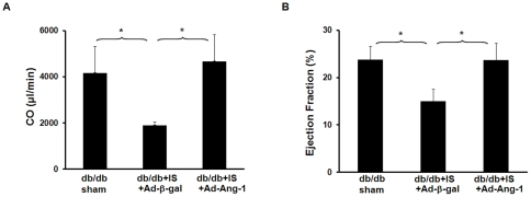
Hematopoietic progenitor CD133(+)/c-kit(+) cells have been shown to be involved in myocardial healing following myocardial infarction (MI). Previously we demonstrated that angiopoietin-1(Ang-1) is beneficial in the repair of diabetic infarcted hearts. We now investigate whether Ang-1 affects CD133(+)/c-kit(+) cell recruitment to the infarcted myocardium thereby mediating cardiac repair in type II (db/db) diabetic mice. db/db mice were administered either adenovirus Ang-1 (Ad-Ang-1) or Ad-β-gal systemically immediately after ligation of the left anterior descending coronary artery (LAD). Overexpression of Ang-1 resulted in a significant increase in CXCR-4/SDF-1α expression and promoted CD133(+)/c-kit(+), CD133(+)/CXCR-4(+) and CD133(+)/SDF-1α(+) cell recruitment into ischemic hearts. Overexpression of Ang-1 led to significant increases in number of CD31(+) and smooth muscle-like cells and VEGF expression in bone marrow (BM). This was accompanied by significant decreases in cardiac apoptosis and fibrosis and an increase in myocardial capillary density. Ang-1 also upregulated Jagged-1, Notch3 and apelin expression followed by increases in arteriole formation in the infarcted myocardium. Furthermore, overexpression of Ang-1 resulted in a significant improvement of cardiac functional recovery after 14 days of ischemia. Our data strongly suggest that Ang-1 attenuates cardiac apoptosis and promotes cardiac repair by a mechanism involving in promoting CD133(+)/c-kit(+) cells and angiogenesis in diabetic db/db mouse infarcted hearts.


Conflict of interest statement

Competing Interests: The authors have declared that no competing interests exist.

Figures

Figure 1
Figure 1. Expression and localization of Ang-1 in the diabetic db/db mouse hearts and bone marrow.
(A) Western blot analysis demonstrating that systemic administration of Ad-Ang-1 (1×10⁹ PFU) resulted in overexpression of Ang-1 in the db/db mouse hearts and bone marrow at day 1 compared to mice receiving Ad-β-gal. (B) Ang-1 was stained with an Ang-1 antibody-labeled with FITC (green, 40X) and nuclei were stained with DAPI (blue, 40X). Merged images show Ang-1 expression in Ad-Ang-1 treated db/db mouse hearts and bone marrow (right panel), but not in Ad-β-gal treated db/db mouse hearts and bone marrow (left panel). (C) Ang-1 protein was localized at smooth muscle cell (SMC) and endothelial cell (EC) in heart tissue. Ang-1 expression was imunostained with Texas Red (red). Endothelial cells were stained with LB4 (green). Merged image revealed that Ang-1 was localized to EC (left panel). Ang-1 was stained with Ang-1 linked to FITC (green). SMCs were stained with SMA (Red) and nuclei were stained with DAPI counterstaining (blue). Merged image showed that Ang-1was localized to SMC (right panel). (D) Western blot analysis demonstrating that systemic administration of Ad-Ang-1 (1×10⁷ –1×10⁹ PFU) resulted in overexpression of Ang-1 in the mouse hearts and bone marrow in a dose-dependent manner.
Figure 2
Figure 2. Ang-1 promotes hematopoietic stem cell recruitment into the ischemic hearts of db/db mouse.
A. Recruitment of CD133+ in db/db mouse hearts subjected to myocardial ischemia at 24 hours. The hematopoietic stem cells were stained with a CD133 antibody (green, 40X). The nuclei were stained by DAPI (blue, 40X). B: Quantitative analysis of CD133+ cells demonstrating that the number of CD133+ cells was significantly increased in the Ad-Ang-1 treated db/db mice (n = 6) compared to that of Ad-β-gal-treated db/db mice (n = 9). CD133+ cells are expressed as the total number per square mm. Sham control (n = 3). All data represent mean ± SD; *p<0.05. ND =  not detected. C. Co-localization of CD133 with c-kit in db/db mouse infarcted hearts at 14 days of MI. CD133+ cells (green); c-kit+ cells (red) and nuclei were stained by DAPI (blue, 40X). C-kit+ cells were recruited into diabetic db/db mouse infarcted hearts in the Ad-Ang-1 treated db/db mice. Merged images further show that CD133+ cells were co-localized with c-kit+ cells. No c-kit+ cells were seen in the Ad-β-gal-treated db/db mice. No specific staining was observed in the sham control group. n = 3 mice each group. D. Recruitment of hematopoietic CD45+ cells in db/db mouse hearts subjected to myocardial ischemia at 24 hours. The hematopoietic cells were stained with a CD45 antibody (red, 40X). The endothelial cells were stained with a Tie-2 antibody (green). The nuclei were stained by DAPI (blue, 40X). No specific staining was observed in the sham control group. E: Quantitative analysis of CD45+ cells demonstrating that the number of CD45+ cells was significantly increased in the Ad-Ang-1 treated db/db mice (n = 6) compared to that of Ad-β-gal-treated db/db mice (n = 9). CD45+ cells are expressed as the total number per square mm. Sham control (n = 3). All data represent mean ± SD; *p<0.05. ND =  not detected.
Figure 3
Figure 3. Ang-1 upregulates SDF-1α and CXCR-4 expression in the ischemic hearts of db/db mouse.
A: Expression of CXCR-4 in db/db mouse hearts subjected to myocardial ischemia at 24 hours. CXCR-4 positive cell (red) and nuclei were stained by DAPI (blue). B: Quantitative analysis showing that the number of CXCR-4+ cells was significantly increased in the Ad-Ang-1 treated db/db mice (n = 6) compared to that of Ad-β-gal-treated db/db mice (n = 9). CXCR-4+ cells are expressed as the total number per square mm. Sham control (n = 3). All data represent mean ± SD; *p<0.05. ND =  not detected. C: Western blot and densitometric analysis of myocardial CXCR-4 expression showing that overexpression of Ang-1 significantly increased CXCR-4 expression in db/db mice compared to that of Ad-β-gal-treated db/db mice. All data represent mean ± SD (n = 4 mice); *p<0.05. D. Western blot and densitometric analysis of myocardial SDF-1α expression. Overexpression of Ang-1 significantly increased SDF-1α expression in db/db mice compared to that of Ad-β-gal-treated db/db mice. All data represent mean ± SD (n = 4 mice); *p<0.05. E. Co-localization of CD133 with CXCR-4 and CD133 with SDF-1α in db/db mouse infarcted hearts. CD133+ cells (green); CXCR-4+ or SDF-1α+ cells (red) and nuclei were stained by DAPI (blue, 40X). Merged images show that CD133+ cells co-localized with both CXCR-4 (upper panel) and SDF-1α (bottom panel) in the Ad-Ang-1 treated db/db mice, but little co-localization was seen in the Ad-β-gal-treated db/db mice. No specific staining was observed in the sham control group.
Figure 4
Figure 4. Ang-1 attenuates myocardial/endothelial apoptosis, and reduces cardiac fibrosis and hypertrophy in db/db mouse.
A. TUNEL-stained heart sections from sham control db/db mouse, db/db mouse treated with Ad-β-gal or Ad-Ang-1 at 24 hours and 14 days of MI. Myocardial and endothelial apoptotic cells in the infarcted area of the ischemic hearts were identified by TUNEL staining (green) and TUNEL/vWF (red) positive staining, and total nuclei by DAPI counterstaining (blue). B and C. Quantitative analysis of apoptotic cells in sham control db/db mouse and infarcted area of db/db mouse treated with Ad-β-gal or Ad-Ang-1 at 24 hours and 14 days after ischemia. TUNEL or TUNEL/vWF positive cells are expressed as the total number of per mm2. Apoptotic cells were significantly decreased in Ad-Ang-1 compared to Ad-β-gal mice. All data represent mean ± SD (n = 4 mice); *p<0.05. D. Representative images and quantitative analysis of apoptotic cells in the remote area of infarction of db/db mouse at 24 hours after ischemia. Apoptotic cells were significantly decreased in Ad-Ang-1 treated db/db mouse hearts (n = 5) compared to Ad-β-gal treated db/db mice (n = 7). All data represent mean ± SD; sham control n = 5, *p<0.05. E. Representative images of TUNEL and SMA-stained heart sections from sham control db/db mouse, db/db mouse treated with Ad-β-gal or Ad-Ang-1 at 24 hours and 14 days of MI. Myocardial and SMCs apoptotic cells in the infarcted area of the ischemic hearts were identified by TUNEL (green) and SMA (red) positive staining, and total nuclei by DAPI counterstaining (blue). F. Representative images of cardiac fibrosis formation and quantitative analysis of cardiac fibrosis area in db/db mice treated with Ad-β-gal or Ad-Ang-1 stained by Masson’s trichrome. Ad-Ang-1 significantly attenuated area of cardiac fibrosis in db/db mice compared to Ad-β-gal mice. All data represent mean ± SD (n = 5 mice); *p<0.05. G. Heart weight/body weight (HW/BW) ratio in db/db and Ad-Ang-1-treated db/db mouse hearts at 14 days after MI. Treatment with Ad-Ang-1 significantly attenuated cardiac hypertrophy in db/db mouse hearts (n = 5) compared to Ad-β-gal mice (n = 4). All data represent mean ± SD; *p<0.05.
Figure 5
Figure 5. Ang-1 increases CD31+ cells and VEGF expression in the BM and promotes capillary formation in db/db mouse hearts.
A. Representative images showing that BM cells differentiate into CD31+ cells in Ad-β-gal treated db/db mice subjected to myocardial ischemia for 24 hours (left panel) and in the Ad-Ang-1treated db/db mice (right panel). B. Quantitative analysis of BM differentiation into CD31+ after myocardial ischemia. The number of BM cells that differentiate into CD31+ was significantly increased in the Ad-Ang-1treated db/db mice compared to the Ad-β-gal treated db/db mice. There was not difference between sham control and ischemic group. All data represent mean ± SD (n = 3 mice); *p<0.05. C. Western blot densitometric analysis of VEGF expression revealed that systemic delivery of Ad-Ang-1 resulted in a significant increase in VEGF expression in the BM of db/db mouse compared to the Ad-β-gal db/db treated mice. All data represent mean ± SD (n = 3 mice); *p<0.05. D and E. Representative images and quantitative analysis showing that treatment with Ad-Ang-1 significantly increased capillary formation in db/db mice subjected to ischemia compared to Ad-â-gal-treated db/db mice. All data represent mean ± SD (n = 7 mice); *p<0.05.
Figure 6
Figure 6. Ang-1 increases number of SMA+ cells in the db/db mouse BM and hearts.
A. Representative images showing that BM cells differentiate into vascular smooth muscle-like (SMA+) cells in Ad-β-gal treated db/db mice subjected to MI for 24 hours (left panel) and in the Ad-Ang-1treated db/db mice (right panel). B. Quantitative analysis showing a significant increase in number of SMA+ cells after myocardial ischemia for 24 hours following Ad-Ang-1 compared to Ad-β-gal in db/db mice subjected to MI. All data represent mean ± SD (n = 3 mice); *p<0.05. C. Representative images of myocardial VSMC in infarcted areas in db/db+IS+Ad-β-gal and db/db+IS+Ad-Ang-l mice stained by Ang-1+/SMA+ cells. Ang-1 was stained with monoclonal anti-angiopoietin-1 linked to FITC (green, 40X). VSMCs were stained with smooth muscle actin (SMA, Red, 40X) and nuclei were stained with DAPI counterstaining (blue, 40x). Merged image showed that over-expressed Ang-1 co-localized with VSMC. Top and middle panel: representative images showing that systemic delivery of Ad-Ang-1 led to a dramatic increase in the number of Ang-1+/SMA+ cells in db/db mouse subjected to myocardial ischemia for 24 hours compared to Ad-β-gal in db/db mice. Bottom panel: representative images showing the newly formed arteriole (Ang-1+/ SMA+) in the Ad-Ang-1 treated db/db mouse heart after ischemia for 14 days.
Figure 7
Figure 7. Ang-1 upregulates Jagged1, Notch3 and apelin expression in db/db mouse hearts.
A. Western blot densitometry analysis of myocardial Jagged 1 expression showing that systemic delivery of Ad-Ang-1 resulted in a significant increase in Jagged 1 expression in the db/db mice subjected to myocardial ischemia for 24 hours compare to the db/db mice treated with Ad-β-gal. All data represent mean ± SD (n = 4 mice); *p<0.05. B. Western blot densitometric analysis of myocardial Notch3 expression revealing that systemic delivery of Ad-Ang-1 (n = 5) resulted in a significant increase in Notch3 expression in db/db mice subjected to myocardial ischemia for 24 hours compare to the db/db mice treated with Ad-β-gal (n = 4). All data represent mean ± SD; *p<0.05. C. Immunofluorescence microscopy showing expression of Notch3 in the vascular wall of db/db mice infarcted hearts treated with Ad-Ang-1. Merged images showed that Notch3 expression was expressed in newly formed arteriole of the Ad-Ang-1 treated db/db mice (right panel), but not the Ad-β-gal-treated db/db mice (left panel). Notch3 was stained with Notch antibody conjugated to FITC (green, 40X) and nuclei were stained by DAPI (blue). No specific staining was observed in the sham control group. D. Western blot densitometry analysis showing that apelin expression was significantly increased in the db/db mice treated with Ad-Ang-1 compare to the Ad-β-gal treated db/db mice at 24 hours of ischemia. All data represent mean ± SD (n = 4 mice); *p<0.05. E. APLNR expression was increased in the Ad-Ang-1 treated db/db mice compare to the Ad-β-gal treated db/db mice at 24 hours of ischemia, but did not achieve a statistical significantly difference. All data represent mean ± SD (n = 4 mice); p>0.05.
Figure 8
Figure 8. Overexpression of Ang-1 improves cardiac functional recovery of post-MI in db/db mice.
A. Cardiac output (CO) was significantly improved in the db/db mice treated with Ad-Ang-1 (n = 5) compare to the Ad-β-gal treated db/db mice (n = 4) at 14 days of ischemia. Sham control (n = 4). All data represent mean ± SD; *p<0.05. B. Myocardial ejection fraction (EF%) was significantly increased in the db/db mice treated with Ad-Ang-1 (n = 5) compare to the Ad-β-gal treated db/db mice (n = 4) at 14 days of ischemia. Sham control (n = 4). All data represent mean ± SD; *p<0.05.

Similar articles

Cited by

References

    1. Suri C, Jones PF, Patan S, Bartunkova S, Maisonpierre PC, et al. Requisite role of angiopoietin-1, a ligand for the TIE2 receptor, during embryonic angiogenesis. Cell. 1996;87:1171–1180. - PubMed
    1. Papapetropoulos A, Fulton D, Mahboubi K, Kalb RG, O’Connor DS, et al. Angiopoietin-1 inhibits endothelial cell apoptosis via the Akt/survivin pathway. J Biol Chem. 2000;275:9102–9105. - PubMed
    1. Joussen AM, Poulaki V, Tsujikawa A, Qin W, Qaum T, et al. Suppression of diabetic retinopathy with angiopoietin-1. Am J Pathol. 2002;160:1683–1693. - PMC - PubMed
    1. Chen JX, Stinnett A. Ang-1 gene therapy inhibits hypoxia-inducible factor-1alpha (HIF-1alpha)-prolyl-4-hydroxylase-2, stabilizes HIF-1alpha expression, and normalizes immature vasculature in db/db mice. Diabetes. 2008;57:3335–3343. - PMC - PubMed
    1. Isner JM, Asahara T. Angiogenesis and vasculogenesis as therapeutic strategies for postnatal neovascularization. J Clin Invest. 1999;103:1231–1236. - PMC - PubMed

Publication types

MeSH terms

Substances