. 2012;7(4):e35924.
doi: 10.1371/journal.pone.0035924. Epub 2012 Apr 30.

The HIV matrix protein p17 subverts nuclear receptors expression and induces a STAT1-dependent proinflammatory phenotype in monocytes

Affiliations

The HIV matrix protein p17 subverts nuclear receptors expression and induces a STAT1-dependent proinflammatory phenotype in monocytes

Barbara Renga et al. PLoS One. 2012.

Abstract

Background: Long-term remission of HIV-1 disease can be readily achieved by combinations of highly effective antiretroviral therapy (HAART). However, a residual persistent immune activation caused by circulating non infectious particles or viral proteins is observed under HAART and might contribute to an higher risk of non-AIDS pathologies and death in HIV infected persons. A sustained immune activation supports lipid dysmetabolism and increased risk for development of accelerated atehrosclerosis and ischemic complication in virologically suppressed HIV-infected persons receiving HAART.

Aim: While several HIV proteins have been identified and characterized for their ability to maintain immune activation, the role of HIV-p17, a matrix protein involved in the viral replication, is still undefined.

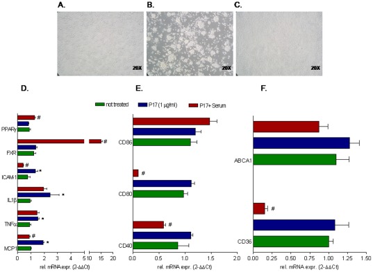
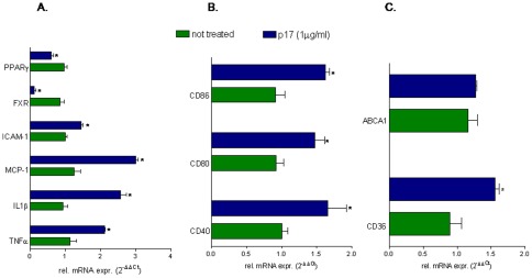
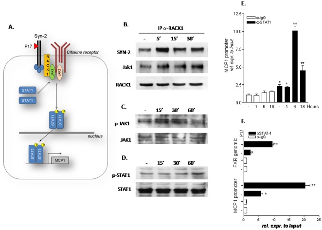
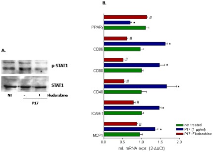
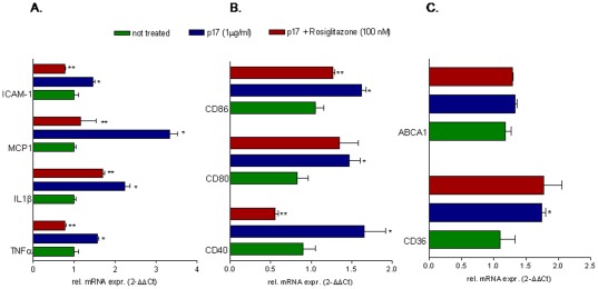
Results: Here, we report that exposure of macrophages to recombinant human p17 induces the expression of proinflammatory and proatherogenic genes (MCP-1, ICAM-1, CD40, CD86 and CD36) while downregulating the expression of nuclear receptors (FXR and PPARγ) that counter-regulate the proinflammatory response and modulate lipid metabolism in these cells. Exposure of macrophage cell lines to p17 activates a signaling pathway mediated by Rack-1/Jak-1/STAT-1 and causes a promoter-dependent regulation of STAT-1 target genes. These effects are abrogated by sera obtained from HIV-infected persons vaccinated with a p17 peptide. Ligands for FXR and PPARγ counteract the effects of p17.

Conclusions: The results of this study show that HIV p17 highjacks a Rack-1/Jak-1/STAT-1 pathway in macrophages, and that the activation of this pathway leads to a simultaneous dysregulation of immune and metabolic functions. The binding of STAT-1 to specific responsive elements in the promoter of PPARγ and FXR and MCP-1 shifts macrophages toward a pro-atherogenetic phenotype characterized by high levels of expression of the scavenger receptor CD36. The present work identifies p17 as a novel target in HIV therapy and grounds the development of anti-p17 small molecules or vaccines.


Conflict of interest statement

Competing Interests: This study was in part supported by Medestea, Turin, Italy. This does not alter the authors' adherence to all the PLoS ONE policies on sharing data and materials..

Figures

Figure 1
Figure 1. HIV-1 p17 regulates pro-inflammatory, co-stimulatory and pro-atherogenic molecules in CD14-derived PBMC isolated from healthy donors.
CD-14 derived PBMC isolated from healthy donors were stimulated for 18 hours with 1 µg/ml p17 recombinant protein. (A and B) Exposure to p17 drives an activated phenotype in macrophages and causes macrophages adhesion. Magnification 20×. (C) Relative mRNA expression of proinflammatory cytokines TNFα, IL1β, MCP-1, ICAM-1 and nuclear receptors FXR and PPARγ. (D) Relative mRNA expression of co-stimulatory molecules CD40, CD80 and CD86. (E) Relative mRNA expression of proatherogenic genes CD36 and ABCA1. Real-Time analysis was carried out in triplicate and the experiment was repeated twice. *P<0.05 versus not treated cells.
Figure 2
Figure 2. HIV-1 p17 exerts regulatory effects on CD-14 derived PBMC isolated from HIV infected patients.
CD-14 derived PBMC isolated from HIV infected patients vaccinated with an anti-p17 vaccine were stimulated ex vivo with 1 µg/ml p17 for 18 hours with or without the serum of the same patients (diluted 1∶100 in medium culture). (A–C) Activation of CD-14 derived PBMC caused by p17 (b) was reserved by p17 immune-neutralization with anti-p17 serum. Magnification 20×. (D) Relative mRNA expression of proinflammatory cytokines TNFα, IL1β, MCP-1, ICAM-1 and nuclear receptors FXR and PPARγ. (E) Relative mRNA expression of co-stimulatory molecules CD40, CD80 and CD86. (F) Relative mRNA expression of proatherogenic genes CD36 and ABCA1. Real-Time analysis was carried out in triplicate and the experiment was repeated twice. *P<0.05 versus not treated cells. #P<0.05 versus p17 stimulated cells.
Figure 3
Figure 3. Stimulation of THP-1 cells with p17 causes reciprocal regulation of genes involved in immune function and lipid metabolism.
THP-1 cells were stimulated with 1 µg/ml p17 recombinant protein for 18 hours. (A) Relative mRNA expression of proinflammatory mediators TNFα, IL1β, MCP-1, ICAM-1 and nuclear receptors FXR and PPARγ. (B) Relative mRNA expression of co-stimulatory molecules CD40, CD80 and CD86. (C) Relative mRNA expression of proatherogenic genes CD36 and ABCA1. Real-Time analysis was carried out in triplicate and the experiment was repeated twice. *P<0.05 versus not treated cells.
Figure 4
Figure 4. p17 signals through RACK-1/Jak-1/STAT-1 pathway in THP-1 cells.
(A) Schematic diagram showing putative interaction of p17 with syndecan-2 and the activation of RACK-1/Jak-1/STAT-1 signal transduction pathway. (B) Co-immuneprecipitation of RACK-1 with syndecan-2 and Jak-1 following stimulation of THP-1 cells with p17 recombinant protein (1 µg/ml) for 5, 15 and 30 minutes. (C) Analysis of Jak-1 protein (total and phosphorylated fraction) by immune blot in THP-1 cells stimulated with p17 (1 µg/ml) for 15, 30 and 60 minutes. (D) Analysis of STAT-1 protein (total and phosphorylated fraction) by immune blot in THP-1 cells stimulated with p17 (1 µg/ml) for 15, 30 and 60 minutes. (E) Chromatin Immunoprecipitation assay carried out in THP-1 cells left untreated or primed with 1 µg/ml p17 for 1, 6 and 18 hours. Real-Time PCR was performed on MCP-1 promoter. (F) Chromatin Immunoprecipitation assay carried out in THP-1 cells left untreated or primed with 1 µg/ml p17 for 18 hours. Real-Time PCR was performed on both MCP-1 promoter and intron-II of FXR gene. Values are normalized relative to input DNA concentration and are expressed relative to those of not treated cells immunoprecipitated with an anti IgG antibody, condition set as 1. Analysis was carried out in triplicate and the experiment was repeated twice. *P<0.05 versus not treated cells. **P<0.05 versus p17 stimulated cells.
Figure 5
Figure 5. An intact STAT-1 signal is required to mediate the biological activity of p17.
THP-1 cells were stimulated for 18 hours with p17 (1 µg/ml) in presence or in absence of a specific STAT-1 inhibitor fludarabine (0.5 µM). At the end of treatments cellular lysates were used for Real-Time or immunoblot analysis. (A) Immunoblot of STAT-1 protein (total and phosphorylated fraction). (B) Relative mRNA expression of MCP-1, ICAM-1, PPARγ, CD40, CD80 and CD86 was expressed relative to not treated cells. Analysis was carried out in triplicate and the experiment was repeated twice. *P<0.05 versus not treated cells. #P<0.05 versus p17 stimulated cells.
Figure 6
Figure 6. FXR agonist, GW-4064, reverses the effects exerted by p17 on THP-1 cells.
THP-1 cells were pre-incubated 2 hours with GW-4064 (1 µM) before administration of p17 (1 µg/ml for 18 hours). Total RNA was extracted and the relative mRNA expression of (A) proinflammatory cytokines and nuclear receptors, (B) co-stimulatory molecules and (C) proatherogenic genes was assayed by Real-Time PCR. Values are expressed relative to not treated cells. Analysis was carried out in triplicate and the experiment was repeated twice. *P<0.05 versus not treated cells. **P<0.05 versus p17 stimulated cells.
Figure 7
Figure 7. The PPARγ agonist, rosiglitazone, differentially regulates p17 biological activities.
THP-1 cells were pre-incubated 2 hours with rosiglitazone (1 µM) before administration of p17 recombinant protein (1 µg/ml for 18 hours). Total RNA was extracted and the relative mRNA expression of (A) proinflammatory cytokines and nuclear receptors, (B) co-stimulatory molecules and (C) proatherogenic genes was analyzed by Real-Time PCR. Values are expressed relative to not treated cells. Analysis was carried out in triplicate and the experiment was repeated twice. *P<0.05 versus not treated cells. **P<0.05 versus p17 stimulated cells.

References

    1. Bukrinsky MI, Haggerty S, Dempsey MP, Sharova N, Adzhubel A, et al. A nuclear localization signal within HIV-1 matrix protein that governs infection of non-dividing cells. Nature. 1993;365:666–669. - PMC - PubMed
    1. Freed EO, Orenstein JM, Buckler-White AJ, Martin MA. Single amino acid changes in the human immunodeficiency virus type 1 matrix protein block virus particle production. J Virol. 1994;68:5311–5320. - PMC - PubMed
    1. Haffar OK, Popov S, Dubrovsky L, Agostini I, Tang H, et al. Two nuclear localization signals in the HIV-1 matrix protein regulate nuclear import of the HIV-1 pre-integration complex. J Mol Biol. 2000;299:359–368. - PubMed
    1. Fiorentini S, Marini E, Caracciolo S, Caruso A. Functions of the HIV-1 matrix protein p17. New Microbiol. 2006;29:1–10. - PubMed
    1. Fiorentini S, Riboldi E, Facchetti F, Avolio M, Fabbri M, et al. HIV-1 matrix protein p17 induces human plasmacytoid dendritic cells to acquire a migratory immature cell phenotype. Proc Natl Acad Sci U S A. 2008;105:3867–3872. - PMC - PubMed

Publication types

MeSH terms

Substances