. 2012;7(4):e36300.
doi: 10.1371/journal.pone.0036300. Epub 2012 Apr 27.

In vitro RNase and nucleic acid binding activities implicate coilin in U snRNA processing

Affiliations

In vitro RNase and nucleic acid binding activities implicate coilin in U snRNA processing

Hanna J Broome et al. PLoS One. 2012.

Abstract

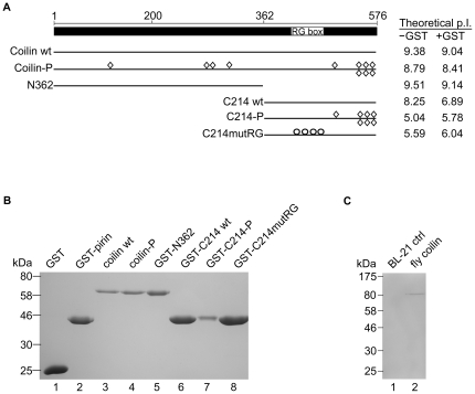
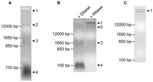
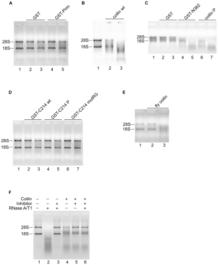
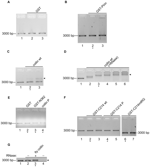
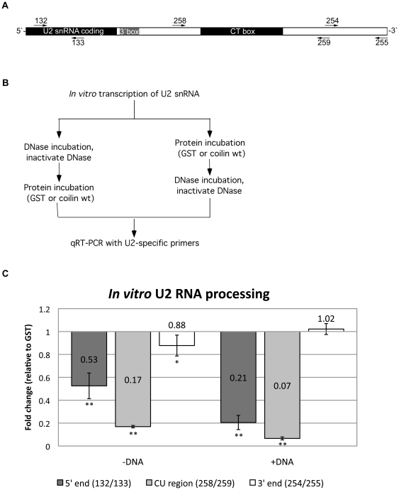
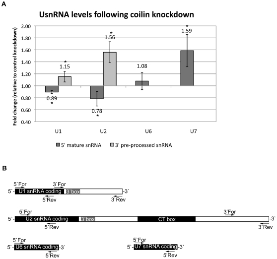
Coilin is known as the marker protein for Cajal bodies (CBs), subnuclear domains important for the biogenesis of small nuclear ribonucleoproteins (snRNPs) which function in pre-mRNA splicing. CBs associate non-randomly with U1 and U2 gene loci, which produce the small nuclear RNA (snRNA) component of the respective snRNP. Despite recognition as the CB marker protein, coilin is primarily nucleoplasmic, and the function of this fraction is not fully characterized. Here we show that coilin binds double stranded DNA and has RNase activity in vitro. U1 and U2 snRNAs undergo a processing event of the primary transcript prior to incorporation in the snRNP. We find that coilin displays RNase activity within the CU region of the U2 snRNA primary transcript in vitro, and that coilin knockdown results in accumulation of the 3' pre-processed U1 and U2 snRNA. These findings present new characteristics of coilin in vitro, and suggest additional functions of the protein in vivo.


Conflict of interest statement

Competing Interests: The authors have declared that no competing interests exist.

Figures

Figure 1
Figure 1. Protein constructs and purification to homogeneity by electro-elution.
A, Schematic of full length human coilin with line diagrams of bacterially expressed constructs below. RG box denotes the 4 Arg-Gly repeat at amino acids 413–420. Construct names are listed to the left and theoretical iso-electric points with and without GST tag are to the right of each construct line diagram. Diamonds represent mutations to Asp or Glu, mimicking phosphorylation by addition of negative charge. Circles represent mutations of the 4 Arg in the RG box to Gly. B, Coomassie stained SDS PAGE with samples from bacterially expressed proteins purified to homogeneity by electro-elution. C, Coomassie stained SDS PAGE with samples from a control BL-21 purification or bacterially expressed fly coilin protein purified to homogeneity by electro-elution.
Figure 2
Figure 2. Coilin purified by electro-elution contains DNA and RNA.
A, 500 ng electro-eluted coilin wt; arrows 1–5 denote distinct nucleic acid species. B, 500 ng electro-eluted coilin wt either DNase I or RNase A/T1 treated for 30 m at 37°C. DNase treated lane contains only RNA species indicated by arrows 3 and 4. RNase treated lanes contains only DNA species indicated by arrows 1 and 5. C, 500 ng electro-eluted coilin wt after 10 days at 4°C.
Figure 3
Figure 3. Purified coilin has RNase activity in its N terminal region.
All reactions, unless indicated, contain either 25 or 100 ng purified electro-eluted protein (left to right) and 500 ng HeLa RNA. After incubation, reactions were loaded into 1% agarose gels containing ethidium bromide. 28S and 18S ribosomal RNA bands are denoted. A control reaction containing RNA but not protein is shown in lane 1 of each panel. Negative control proteins are GST and GST-pirin. A, negative controls GST and GST-pirin. B, coilin wt. C, negative control, GST; coilin constructs GST-N362 and coilin P. D, coilin C terminal constructs. E, D. melanogaster (fly) coilin. F, incubations of 250 ng coilin wt and/or RNase A/T1 cocktail (2.5 pg RNase A and 0.075 pg RNase T1) with 500 ng HeLa RNA with or without 0.1 units RNase inhibitor; + or − signs denote presence or absence of the component listed to the left in each reaction.
Figure 4
Figure 4. Purified coilin binds DNA.
All reactions contain purified electro-eluted protein and 13 ng DNA. After incubation, reactions were loaded into 1% agarose gels containing ethidium bromide. The location of the approximately 3000 bp unbound DNA is indicated. Lane 1 for each gel represents control reactions without protein. A, negative control GST; Lane 2, 0.22 µg; Lane 3, 2.2 µg. B, negative control GST-pirin; Lane 2, 0.2 µg; Lane 3, 2 µg. C, coilin wt; Lane 2, 0.13 µg; Lane 3, 0.5 µg. D, RNase treated coilin wt; Lanes 2–6, 0.1, 0.2, 0.4, 0.6, 0.8 µg. E, negative control GST, 0.29 µg; coilin construct GST-N362, 0.29 µg; coilin P construct, 0.29 µg. F, GST-C214, GST-C214 P and GST-C214mtRG; Lanes 2–3, 0.29 and 0.57 µg; Lanes 4–5, 0.29 and 0.57 µg; Lanes 6–7, 0.2 and 2 µg. G, D. melanogaster coilin either untreated or pre-treated with RNase A/T1; Lane 2, 0.19 µg untreated protein; Lane 3, 0.19 µg RNase treated; Lane 4, 0.19 untreated protein alone; line is drawn to highlight the slight DNA mobility shift. Arrows in C, D, and G indicate the location of a distinct protein/DNA complex, resulting in slower migration than unbound DNA. Asterisk (*) under lanes denotes protein amount included in Table 1 for DNA binding.
Figure 5
Figure 5. Purified coilin cleaves U2 RNA transcript.
A, Diagram of U2 gene repeat region including snRNA coding region and extending ∼630 bp beyond. Primers used for qRT-PCR are denoted; forward primers above and reverse primers below diagram. B, Diagram of protocol for incubations and subsequent analysis of U2 RNA via qRT-PCR. C, graph of relative U2 RNA qRT-PCR product amount following incubation with purified electro-eluted coilin. Values represent fold change of product levels following coilin incubation, normalized to GST incubation set at 1. Error bars represent 1 standard deviation of fold change, n = 9. Statistical analysis performed using a paired Student's Ttest of GST incubated and coilin incubated Ct values. * denotes p<0.03, ** denotes p<0.0005.
Figure 6
Figure 6. Coilin knockdown results in accumulation of primary U snRNA transcripts.
A, relative U snRNA levels in HeLa cells following coilin knockdown. Error bars represent 1 standard deviation of fold change, n = 9. Statistical analysis performed using a paired Student's Ttest of the change in Ct relative to GAPDH between control and coilin knockdown RNA. * denotes p<0.04. C, diagrams of U snRNA genes with locations of primers used for qRT-PCR analysis noted.

References

    1. Morris GE. The Cajal body. Biochim Biophys Acta. 2008;1783:2108–2115. - PubMed
    1. Matera AG, Izaguire-Sierra M, Praveen K, Rajendra TK. Nuclear bodies: random aggregates of sticky proteins or crucibles of macromolecular assembly? Dev Cell. 2009;17:639–647. - PMC - PubMed
    1. Frey MR, Matera AG. Coiled Bodies Contain U7 Small Nuclear RNA and Associate with Specific DNA Sequences in Interphase Cells. Proc Natl Acad Sci USA. 1995;92:5915–5919. - PMC - PubMed
    1. Cuello P, Boyd DC, Dye MJ, Proudfoot NJ, Murphy S. Transcription of the human U2 snRNA genes continues beyond the 3′ box in vivo. Embo J. 1999;18:2867–2877. - PMC - PubMed
    1. Medlin JE, Uguen P, Taylor A, Bentley DL, Murphy S. The C-terminal domain of pol II and a DRB-sensitive kinase are required for 3′ processing of U2 snRNA. Embo J. 2003;22:925–934. - PMC - PubMed

Publication types