. 2012;7(4):e36373.
doi: 10.1371/journal.pone.0036373. Epub 2012 Apr 30.

Globotriaosylsphingosine accumulation and not alpha-galactosidase-A deficiency causes endothelial dysfunction in Fabry disease

Affiliations

Globotriaosylsphingosine accumulation and not alpha-galactosidase-A deficiency causes endothelial dysfunction in Fabry disease

Mehdi Namdar et al. PLoS One. 2012.

Abstract

Background: Fabry disease (FD) is caused by a deficiency of the lysosomal enzyme alpha-galactosidase A (GLA) resulting in the accumulation of globotriaosylsphingosine (Gb3) in a variety of tissues. While GLA deficiency was always considered as the fulcrum of the disease, recent attention shifted towards studying the mechanisms through which Gb3 accumulation in vascular cells leads to endothelial dysfunction and eventually multiorgan failure. In addition to the well-described macrovascular disease, FD is also characterized by abnormalities of microvascular function, which have been demonstrated by measurements of myocardial blood flow and coronary flow reserve. To date, the relative importance of Gb3 accumulation versus GLA deficiency in causing endothelial dysfunction is not fully understood; furthermore, its differential effects on cardiac micro- and macrovascular endothelial cells are not known.

Methods and results: In order to assess the effects of Gb3 accumulation versus GLA deficiency, human macro- and microvascular cardiac endothelial cells (ECs) were incubated with Gb3 or silenced by siRNA to GLA. Gb3 loading caused deregulation of several key endothelial pathways such as eNOS, iNOS, COX-1 and COX-2, while GLA silencing showed no effects. Cardiac microvascular ECs showed a greater susceptibility to Gb3 loading as compared to macrovascular ECs.

Conclusions: Deregulation of key endothelial pathways as observed in FD vasculopathy is likely caused by intracellular Gb3 accumulation rather than deficiency of GLA. Human microvascular ECs, as opposed to macrovascular ECs, seem to be affected earlier and more severely by Gb3 accumulation and this notion may prove fundamental for future progresses in early diagnosis and management of FD patients.


Conflict of interest statement

Competing Interests: The authors have declared that no competing interests exist.

Figures

Figure 1
Figure 1. Gb3 loading and alpha-Galactosidase A gene silencing.
A. Incubation with Gb3 increased intracellular concentration of Gb3, as measured by ELISA, by 2-folds in HMaVECs and HMiVECs (n = 4, p = 0.03 and p = 0.04, respectively, versus untreated control, B. Maximal a-Gal A protein silencing of 70% occurred 48 hrs after transfection of HMaVECs with siRNA 3. Transfection of HMaVECs with Non-targeting (NT)-RNA did not alter endothelial a-Gal A expression. Values are representative of at least 4 different experiments; all blots are normalized to GAPDH expression. C. Intracellular Gb3 concentrations 2 days following siRNA transfection remained unchanged compared to untreated cells.
Figure 2
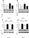
Figure 2. Gb3 impairs eNOS expression in HMiVECs.
A. Gb3 inhibits total eNOS expression in TNF-α stimulated HMiVECs and under basal conditions. Values are indicated as percent of unstimulated control. *p<0.05 vs unstimulated control; **p<0.05 vs TNF-α alone. B. Gb3 inhibits Ser1177 pho eNOS expression in TNF-α stimulated HMiVECs and under basal conditions. Values are indicated as percent of unstimulated control. *p<0.05 vs unstimulated control; **p<0.05 vs TNF-α alone. Values are representative of at least 4 different experiments; all blots are normalized to GAPDH expression. C./D. In all cases expression remained unchanged following GLA gene silencing.
Figure 3
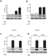
Figure 3. Gb3 enhances iNOS expression in HMiVECs.
A. Gb3 enhances iNOS expression in HMiVECs under basal conditions. TNF-α enhances iNOS expression in HMiVECs. Values are indicated as percent of unstimulated control. *p<0.05 vs unstimulated control. B. TNF-α enhances iNOS expression in HMaVECs. Values are indicated as percent of unstimulated control. *p<0.05 vs unstimulated control; Values are representative of at least 4 different experiments; all blots are normalized to GAPDH expression. C./D. GLA gene silencing had no effect on expression of iNOS.
Figure 4
Figure 4. Gb3 enhances COX-2 expression in HMiVECs.
A. Gb3 enhances COX-2 expression in HMiVECs under basal conditions. TNF-α enhances COX-2 expression in HMiVECs. Values are indicated as percent of unstimulated control. *p<0.05 vs unstimulated control. B. TNF-α enhanced COX-1 expression in both HMiVECs and HMaVECs by 1.5-fold and 1.4-fold, respectively. Values are representative of at least 4 different experiments; all blots are normalized to GAPDH expression. C. GLA gene silencing had no effect on expression of COX-2.
Figure 5
Figure 5. Gb3 enhances VCAM-1 expression in TNF-α stimulated HMiVECs.
A. TNF-α enhances VCAM-1 expression in HMiVECs. Values are indicated as percent of unstimulated control. *p<0.05 vs unstimulated control; Values are representative of at least 4 different experiments; all blots are normalized to GAPDH expression. B. Gb3 enhances VCAM-1 expression in TNF-α stimulated HMaVECs and under basal conditions. Values are indicated as percent of unstimulated control. *p<0.05 vs unstimulated control; **p<0.05 vs TNF-α alone. C./D. GLA gene silencing did not affect expression of VCAM-1.

References

    1. Zarate YA, Hopkin RJ. Fabry's disease. Lancet. 2008;372:1427–1435. - PubMed
    1. Park JL, Whitesall SE, D'Alecy LG, Shu L, Shayman JA. Vascular dysfunction in the alpha-galactosidase A-knockout mouse is an endothelial cell-, plasma membrane-based defect. Clin Exp Pharmacol Physiol. 2008;35:1156–1163. - PMC - PubMed
    1. Elliott PM, Kindler H, Shah JS, Sachdev B, Rimoldi OE, et al. Coronary microvascular dysfunction in male patients with Anderson-Fabry disease and the effect of treatment with alpha galactosidase A. Heart. 2006;92:357–360. - PMC - PubMed
    1. Kalliokoski RJ, Kalliokoski KK, Sundell J, Engblom E, Penttinen M, et al. Impaired myocardial perfusion reserve but preserved peripheral endothelial function in patients with Fabry disease. J Inherit Metab Dis. 2005;28:563–573. - PubMed
    1. Wang RY, Lelis A, Mirocha J, Wilcox WR. Heterozygous Fabry women are not just carriers, but have a significant burden of disease and impaired quality of life. Genet Med. 2007;9:34–45. - PubMed

Publication types

MeSH terms