One patient with double lymphomas: simultaneous gastric MALT lymphoma and ileal diffuse large B-cell lymphoma
- PMID: 22558482
- PMCID: PMC3341684
One patient with double lymphomas: simultaneous gastric MALT lymphoma and ileal diffuse large B-cell lymphoma
Abstract
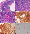
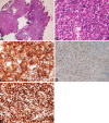
Multiple different lymphomas in a single person are very rare. The author herein reports the case of a 69- year-old Japanese woman with double gastrointestinal lymphoma. The patient presented with epigastralgia. Endoscopic examination revealed erosions and elevation of the gastric body and a large ulcerated tumor of the terminal ileum. Biopsies were obtained from these lesions. The gastric lesion was MALT lymphoma with monocytoid B-cell proliferation and lymphoepithelial lesions. Light chain restriction was present. Helicobacter pylori were present on Giemsa stain. The gastric lesions did not regress despite of therapy, which were confirmed by follow-up biopsy. The ileal lesion was obvious diffuse large B-cell lymphoma. The lesion regressed by chemotherapy. The patient is now alive 3 years after the first presentation.
Keywords: Double lymphoma; gastric MALT lymphoma; ileal diffuse large B-cell lymphoma.
Figures


References
-
- Tang Z, Jing W, Lindeman N, Harris NL, Ferry JA. One patient, two lymphomas: Simultaneous primary gastric marginal zone lymphoma and primary duodenal follicular lymphoma. Arch Pathol Lab Med. 2004;128:1035–1038. - PubMed
-
- Jaffe ES, Harris NL, Stein H, Vardiman JW, editors. WHO Classification of tumour. Pathology and Genetics of tumors of hematopoietic and lymphoid system. Lyon: IARC Press; 2001.
-
- Terada T, Kawaguchi M, Furukawa K, Sekido Y, Osamura Y. Minute mixed ductal-endocrine carcinoma of the pancreas with predominant intraductal growth. Pathol Int. 2002;52:740–746. - PubMed
-
- Terada T, Kawaguchi M. Primary clear cell adenocarcinoma of the peritoneum. Tohoku J Exp Med. 2005;206:271–275. - PubMed
Publication types
MeSH terms
Substances
LinkOut - more resources
Full Text Sources
Medical
