

Review
doi: 10.1021/cr2001965.
Epub 2012 May 4.
Chromophore transformations in red fluorescent proteins
Affiliations
- PMID: 22559232
- PMCID: PMC3394910
- DOI: 10.1021/cr2001965
Item in Clipboard
Review
Chromophore transformations in red fluorescent proteins
Chem Rev.
.
No abstract available
Figures
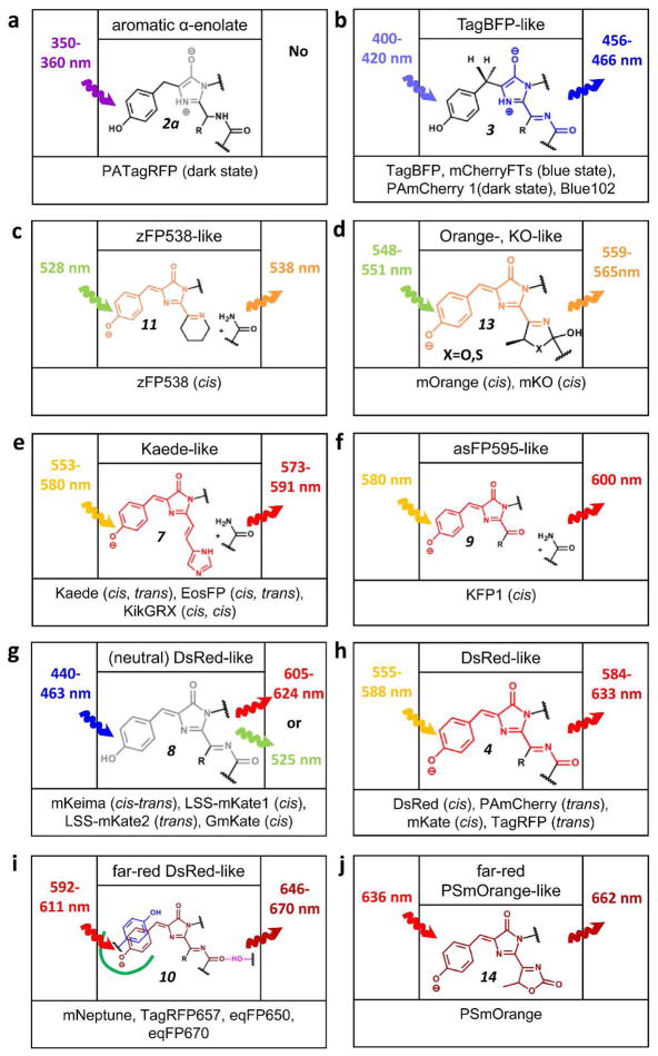
Chromophores in RFPs are shown in an order of their fluorescence emission color (from a through i). Excitation and emission maxima of respective chromophore for indicated FPs are shown on left and right sides, respectively. Configuration of a double bond between the Tyr-ring and imidazolinone is shown in the brackets. For Kaede-like chromophore configuration of the double bond between His-ring and imidazolinone is shown in the second position in the brackets. Numbering of the chromophore structures follows that in Figures 2 and 4. References and PDB ID numbers corresponding to the respective chromophores are listed in Table 1.
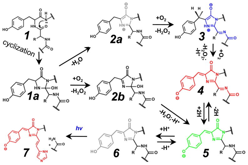
Mechanisms of autocatalytic formation of the chromophores in fluorescent proteins are shown. The color of the chemical structures corresponds to the spectral range of the chromophore fluorescence emission, where the gray color denotes the non-fluorescent state. Many of the indicated steps may also be photo-induced. Chromophores are presented in a cis-isoform. Possible trans-isoforms are not shown. The hv symbol designates UV-light.
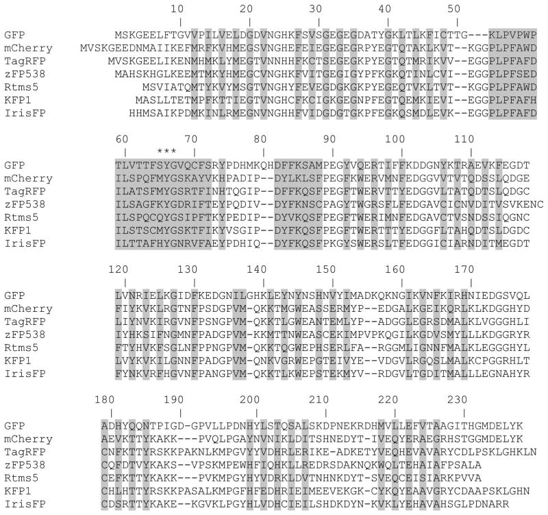
Alignment of amino acid sequences for selected RFPs relative to Aequorea victoria GFP. Alignment numbering follows that of GFP. Residues buried inside of FPs’β-barrels are shaded. Asterisks indicate the residues that form the chromophore.
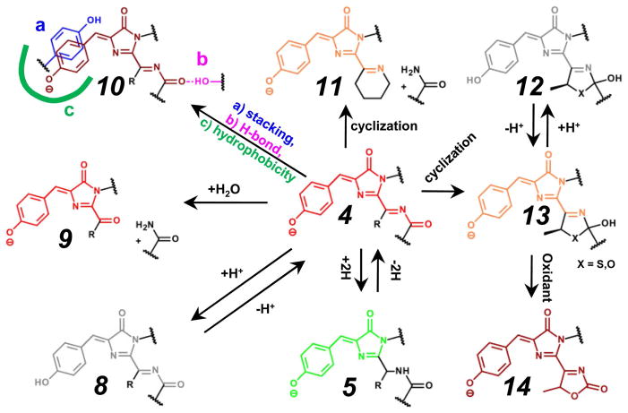
Major autocatalytic modifications of the red chromophore in FPs and their interaction with the immediate surroundings are shown. The color of the chemical structures of chromophores corresponds to the spectral range of the chromophore fluorescence emission, where gray color denotes the non-fluorescent state. Many of the indicated chromophore transformations may also be photo-induced. Chromophores are presented in a cis-form. Possible cis-trans isomerizations are not shown. Transition 4→10a, stacking interactions with tyrosine ring are shown, however, a similar interaction with Phe, Arg, His, or Trp residues are also possible. Transition 4→10c, a green line denotes hydrophobic amino acid’s surrounding of the chromophore. Transition 13→14, an oxidant is molecular oxygen or chemical compound such as potassium ferricyanide or benzoquinone.
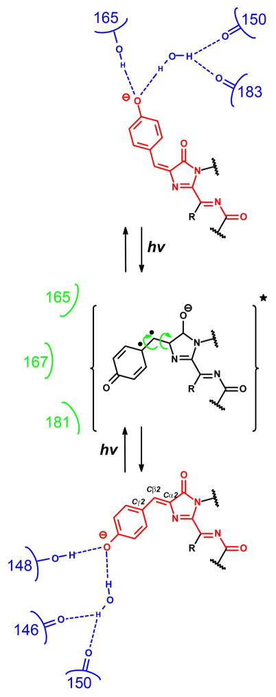
Cis-trans isomerization of the red chromophore in fluorescent proteins induced by light and regulated by surrounding amino acids. The suggested intermediate (data from ref. ) representing chromophore in excited state is shown in brackets. Residues stabilizing chromophore in its cis- or trans-configuration via hydrogen bonds (dash lines) are shown in blue color. Hydroxyl group of the Tyr-ring of the chromophore can be protonated in cis- or trans-configuration of the chromophore. Residues hindering movement of Tyr ring by direct or indirect steric clashing are shown in green color. Electrons on the high-energy orbitals are shown as dots.
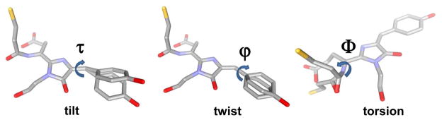
Characteristics of chromophore planarity in terms of tilt (τ) and twist (ϕ) and torsion (Ψ) angles. Tilt and twist angles correspond to rotation of the Tyr-ring of the chromophore around the Cα2-Cβ2 and Cβ2-Cγ2 bonds, respectively. Torsion angle is the dihedral angle defined by atoms X65:Cβ1 - X65:Cα1 - X65:N1 - X64:C, where X65 and X64 denote the residue 65 of the chromophore-forming tripeptide and the residue 64 preceding the chromophore, respectively.
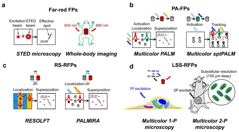
Applications of RFPs in modern fluorescence microscopy techniques. (a) Use of far-red shifted FPs in super-resolution STED microscopy and the whole-body imaging. On the left, doughnut-shaped depletion beam confining excitation beam to an effective spot. On the right, whole-body imaging of frog expressing Katushka far-red FP. (b) Dark-to-red and dark-to-green PA-FPs in multicolor super-resolution PALM and sptPALM microscopy. On the top, photoactivation/photobleaching cycles for PA-RFP and PA-GFP. Red and green Gaussian-shaped curves stand for center localization procedure. Red and green arrows show movement of PA-RFP and PA-GFP particles, respectively. (c) Reversibly switchable FPs (RS-FPs) in RESOLFT and PALMIRA super-resolution STED-based techniques. On the top, photoswitching cycles for KFP and rsCherryRev. Red and green Gaussian-shaped curves stand for center localization procedure. Blue and yellow thick strokes denote grooves of OFF- and ON-light, respectively. (d) Utilization of LSS-RFPs in combination with conventional cyan, green and/or yellow FPs in one- and two-photon imaging of the cell using single wavelength of excitation. On the left, plasma membrane (yellow, CFP), endoplasmic reticulum (cyan, mMiCy), Golgi (green, GFP), microtubules (red, YFP), nucleus (dark blue, dKeima570) and mitochondria (purple, mKeima) visualized by respective FPs. On the right, two-photon imaging of tumor cells in a mouse through optical window; nucleus (red, LSSmKate), cytoplasma (green, GFP) and Golgi (cyan, CFP) are labeled with respective FP. FP molecules are shown as cylinders colored according to their emission colors. Colored waved arrows denote the respective activation/switching, excitation or emission light.
References
Publication types
MeSH terms
Substances
Grants and funding
LinkOut - more resources
Full Text Sources
Other Literature Sources
