

Increasing the oscillation frequency of strong magnetic fields above 101 kHz significantly raises peripheral nerve excitation thresholds
- PMID: 22559628
- PMCID: PMC3344882
- DOI: 10.1118/1.3702775
Increasing the oscillation frequency of strong magnetic fields above 101 kHz significantly raises peripheral nerve excitation thresholds
Abstract
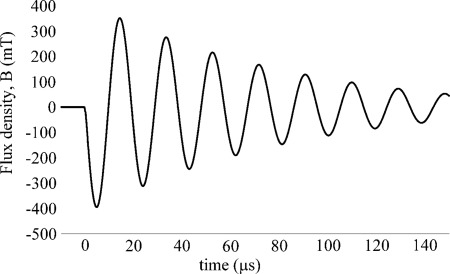
Purpose: A time-varying magnetic field can cause unpleasant peripheral nerve stimulation (PNS) when the maximum excursion of the magnetic field (ΔB) is above a frequency-dependent threshold level [P. Mansfield and P. R. Harvey, Magn. Reson. Med. 29, 746-758 (1993)]. Clinical and research magnetic resonance imaging (MRI) gradient systems have been designed to avoid such bioeffects by adhering to regulations and guidelines established on the basis of clinical trials. Those trials, generally employing sinusoidal waveforms, tested human responses to magnetic fields at frequencies between 0.5 and 10 kHz [W. Irnich and F. Schmitt, Magn. Reson. Med. 33, 619-623 (1995), T. F. Budinger et al., J. Comput. Assist. Tomogr. 15, 909-914 (1991), and D. J. Schaefer et al., J. Magn. Reson. Imaging 12, 20-29 (2000)]. PNS thresholds for frequencies higher than 10 kHz had been extrapolated, using physiological models [J. P. Reilly et al., IEEE Trans. Biomed. Eng. BME-32(12), 1001-1011 (1985)]. The present study provides experimental data on human PNS thresholds to oscillating magnetic field stimulation from 2 to 183 kHz. Sinusoidal waveforms were employed for several reasons: (1) to facilitate comparison with earlier reports that used sine waves, (2) because prior designers of fast gradient hardware for generalized waveforms (e.g., including trapezoidal pulses) have employed quarter-sine-wave resonant circuits to reduce the rise- and fall-times of pulse waveforms, and (3) because sinusoids are often used in fast pulse sequences (e.g., spiral scans) [S. Nowak, U.S. patent 5,245,287 (14 September 1993) and K. F. King and D. J. Schaefer, J. Magn. Reson. Imaging 12, 164-170 (2000)].
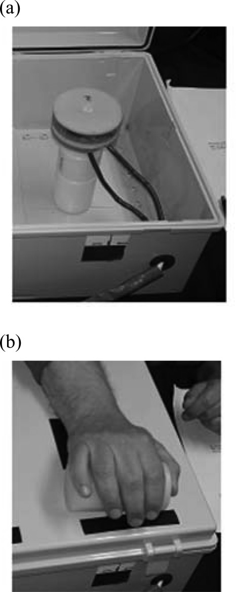
Methods: An IRB-approved prospective clinical trial was performed, involving 26 adults, in which one wrist was exposed to decaying sinusoidal magnetic field pulses at frequencies from 2 to 183 kHz and amplitudes up to 0.4 T. Sham exposures (i.e., with no magnetic fields) were applied to all subjects.
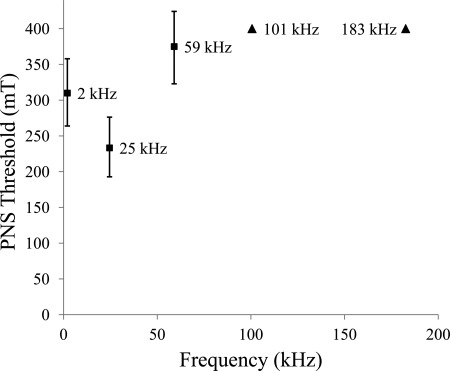
Results: For 0.4 T pulses at 2, 25, 59, 101, and 183 kHz, stimulation was reported by 22 (84.6%), 24 (92.3%), 15 (57.7%), 2 (7.7%), and 1 (3.8%) subjects, respectively.
Conclusions: The probability of PNS due to brief biphasic time-varying sinusoidal magnetic fields with magnetic excursions up to 0.4 T is shown to decrease significantly at and above 101 kHz. This phenomenon may have particular uses in dynamic scenarios (e.g., cardiac imaging) and in studying processes with short decay times (e.g., electron paramagnetic resonance imaging, bone and solids imaging). The study suggests the possibility of new designs for human and preclinical MRI systems that may be useful in clinical practice and scientific research.
Figures

References
-
- Reilly J. P. and Diamant A. M., “Spatially extended nonlinear node (SENN) model of electrostimulation of myelinated nerve,” available at http://www.artechhouse.com/static/reslib/reilly/reilly.html.
-
- Reilly J. P., Applied Bioelectricity (Springer, New York, 1998).
-
- Reilly J. P. and Diamant A. M., Electrostimulation (Artech House, Boston, 2011).
Publication types
MeSH terms
Grants and funding
LinkOut - more resources
Full Text Sources
Other Literature Sources
