

Review
doi: 10.1016/j.smim.2012.04.006.
Epub 2012 May 2.
Genetic regulation of thymocyte progenitor aging
Affiliations
- PMID: 22559986
- PMCID: PMC3415574
- DOI: 10.1016/j.smim.2012.04.006
Item in Clipboard
Review
Genetic regulation of thymocyte progenitor aging
Semin Immunol.
2012 Oct.
Abstract
The number of T cell progenitors is significantly reduced in the involuted thymus, and the growth and developmental potential of the few cells that are present is severely attenuated. This review provides an overview of how aging affects T cell precursors before and following entry into the thymus and discusses the age-related genetic changes that may occur in them. Finally, interventions that rejuvenate thymopoiesis in the elderly by targeting T cell progenitors are discussed.
Copyright © 2012 Elsevier Ltd. All rights reserved.
Figures
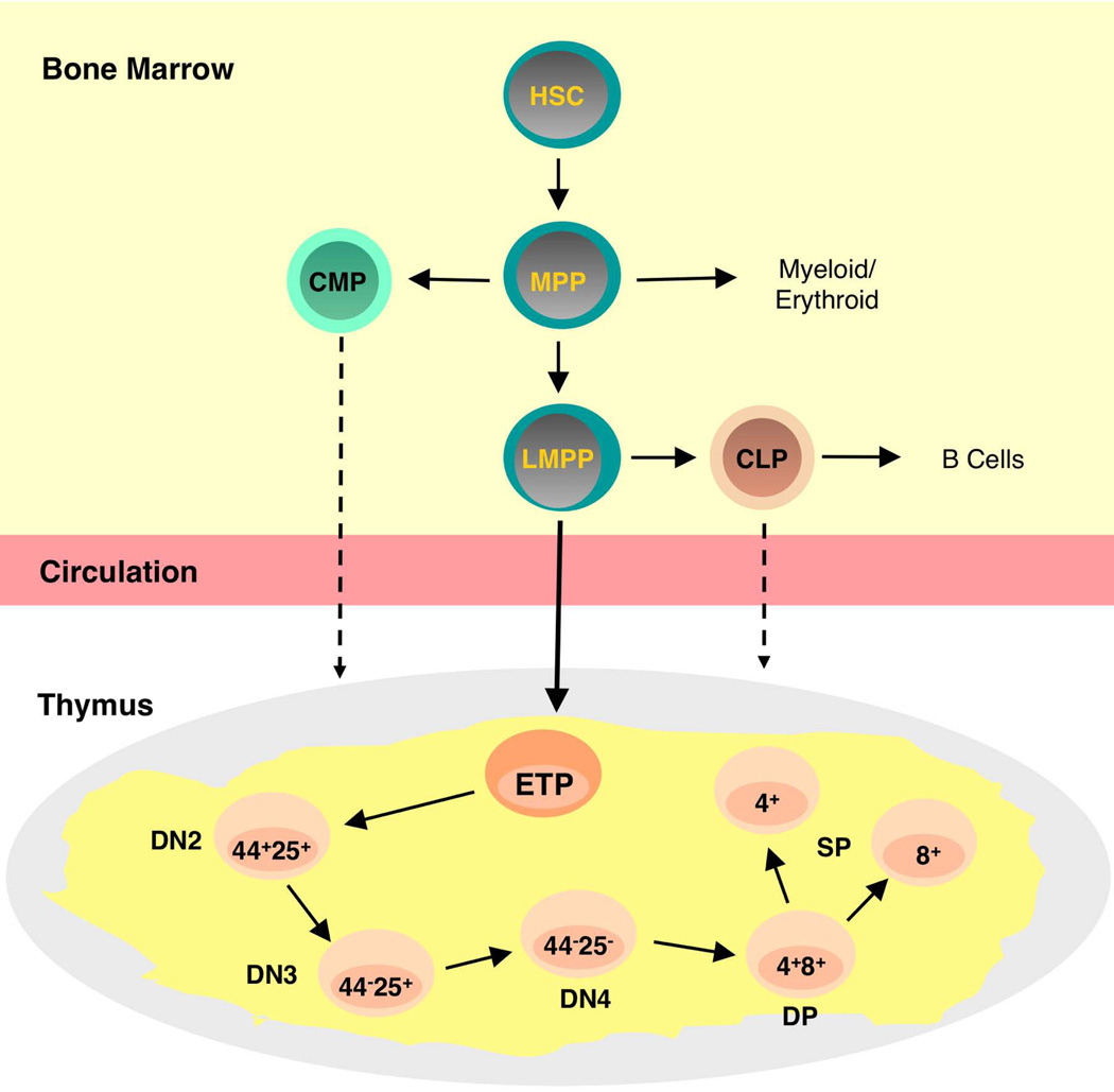
T cell progenitors in the bone marrow and thymus. Hematopoietic stem cells (HSCs) in the bone marrow generate Multipotential Progenitors (MPP) capable of producing lymphoid and myeloid progeny. Lymphoid Primed Multipotential Progenitors (LMPP) have lost most myeloid and erythroid potential and are candidate thymus seeding cells as shown by the solid arrow. Early T Lineage Progenitors (ETP) are the most immature intrathymic progenitor and are defined by their lineage negative CD44high CD25− CD117+ phenotype. ETP then mature into double negative (DN) stages defined by differential expression of CD44 and/or CD25. DN4 cells express the T cell receptor at low levels and then mature into CD4+CD8+ double positive (DP) thymocytes from which CD4 or CD8 single positive (SP) thymocytes derive. Note that other cells in addition to LMPPs may under some circumstances seed the thymus. These could include HSCs, CLPs, and even CMPs. The latter cells have recently been demonstrated to have residual T cell potential.
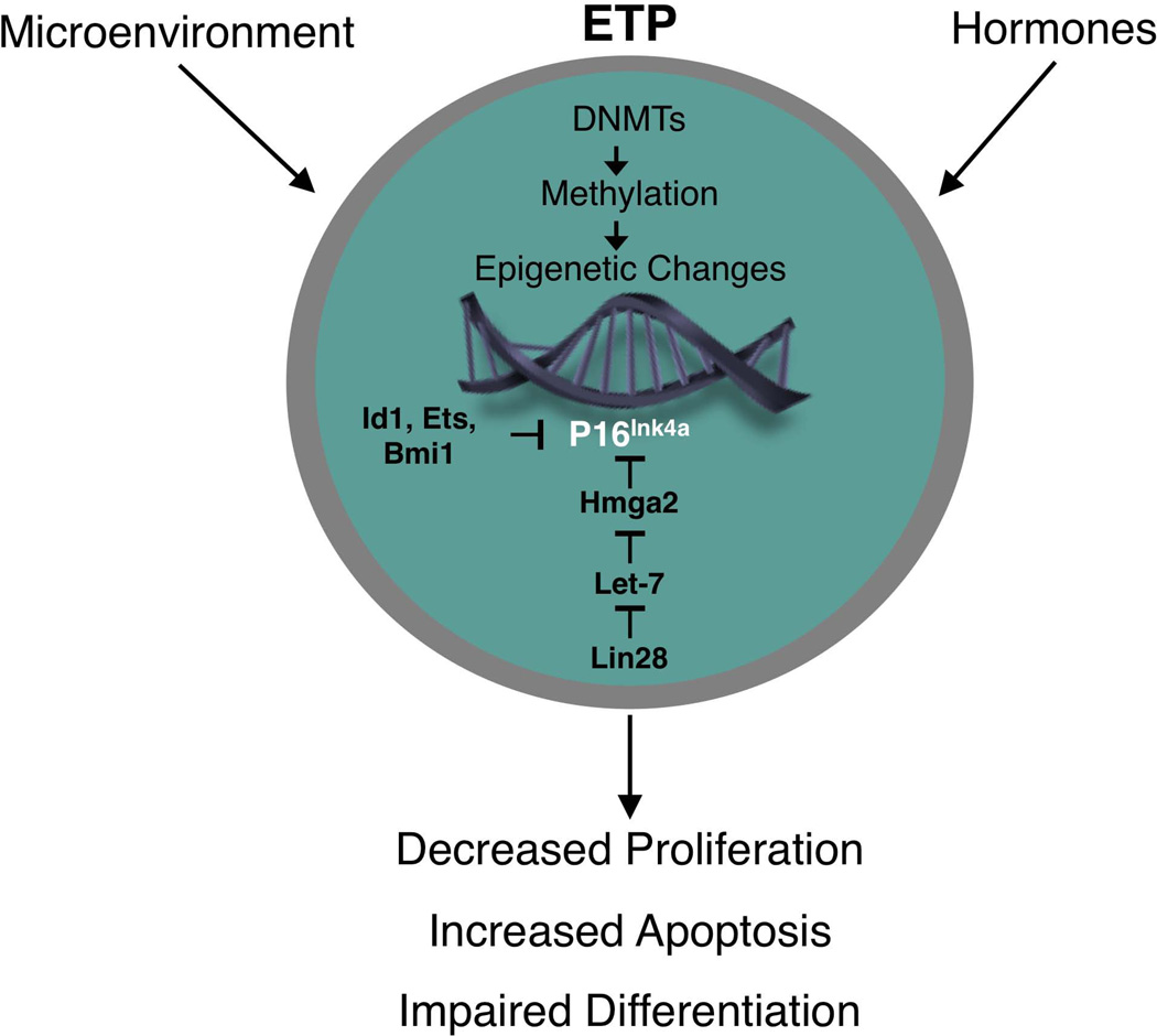
Proposed changes in gene expression in aging ETP. Changes in expression of one or more DNA methyltransferases (DNMTs), as a result of cell intrinsic or extracellular influences, may cause epigenetic modifications that affect the expression of genes that regulate ETP growth, differentiation, and/or survival. Our preliminary data indicate that expression of p16INK4a is increased in ETP with age and is a factor that contributes to their reduced proliferation. A complex network regulates expression of p16INK4a in cells. For example, various factors that include Id1, Ets1, Ets2, Bmi1, and Hmga2 can suppress p16INK4a expression. Hmga2 in turn is under control of multiple upstream factors that include Lin 28 and the Let-7b microRNA. The precise factors that regulate p16INK4a expression in ETP have not been defined.
Similar articles
-
Autonomous and extrinsic regulation of thymopoiesis in human immune system (HIS) mice.Eur J Immunol. 2011 Oct;41(10):2883-93. doi: 10.1002/eji.201141586. Epub 2011 Aug 30. Eur J Immunol. 2011. PMID: 21739431
-
IL-18 acts in synergy with IL-7 to promote ex vivo expansion of T lymphoid progenitor cells.J Immunol. 2015 Apr 15;194(8):3820-8. doi: 10.4049/jimmunol.1301542. Epub 2015 Mar 16. J Immunol. 2015. PMID: 25780034 Free PMC article.
-
Fibroblast growth factor-7 partially reverses murine thymocyte progenitor aging by repression of Ink4a.Blood. 2012 Jun 14;119(24):5715-21. doi: 10.1182/blood-2011-12-400002. Epub 2012 May 3. Blood. 2012. PMID: 22555975 Free PMC article.
-
Thymus-bound: the many features of T cell progenitors.Front Biosci (Schol Ed). 2011 Jun 1;3(3):961-9. doi: 10.2741/200. Front Biosci (Schol Ed). 2011. PMID: 21622245 Review.
-
Thymocyte migration and emigration.Immunol Lett. 2024 Jun;267:106861. doi: 10.1016/j.imlet.2024.106861. Epub 2024 Apr 30. Immunol Lett. 2024. PMID: 38697225 Review.
Cited by
-
Defective respiration and one-carbon metabolism contribute to impaired naïve T cell activation in aged mice.Proc Natl Acad Sci U S A. 2018 Dec 26;115(52):13347-13352. doi: 10.1073/pnas.1804149115. Epub 2018 Dec 10. Proc Natl Acad Sci U S A. 2018. PMID: 30530686 Free PMC article.
-
Lymphocyte generation and population homeostasis throughout life.Semin Hematol. 2017 Jan;54(1):33-38. doi: 10.1053/j.seminhematol.2016.10.003. Epub 2016 Oct 19. Semin Hematol. 2017. PMID: 28088985 Free PMC article. Review.
-
Global transcriptional profiling reveals distinct functions of thymic stromal subsets and age-related changes during thymic involution.Cell Rep. 2014 Oct 9;9(1):402-415. doi: 10.1016/j.celrep.2014.08.070. Epub 2014 Oct 2. Cell Rep. 2014. PMID: 25284794 Free PMC article.
-
The Confluence of Sex Hormones and Aging on Immunity.Front Immunol. 2018 Jun 4;9:1269. doi: 10.3389/fimmu.2018.01269. eCollection 2018. Front Immunol. 2018. PMID: 29915601 Free PMC article. Review.
-
Dysregulation of Systemic Immunity in Aging and Dementia.Front Cell Neurosci. 2021 Jun 22;15:652111. doi: 10.3389/fncel.2021.652111. eCollection 2021. Front Cell Neurosci. 2021. PMID: 34239415 Free PMC article. Review.
References
-
- Linton PJ, Dorshkind K. Age-related changes in lymphocyte development and function. Nat Immunol. 2004;5:133–139. - PubMed
-
- Andrew D, Aspinall R. Thymic atrophy in the mouse is a soluble problem of the thymic environment. Vaccine. 2000;18:1629–1637. - PubMed
-
- Mackall CL, Punt JA, Morgan P, Farr AG, Gress RE. Thymic function in young/old chimeras: substantial thymic T cell regenerative capacity despite irreversible age-associated thymic involution. Eur J Immunol. 1998;28:1886–1893. - PubMed
-
- Sharp A, Kukulansky T, Globerson A. In vitro analysis of age-related changes in the developmental potential of bone marrow thymocyte progenitors. Eur J Immunol. 1990;20:2541–2546. - PubMed
Publication types
MeSH terms
Grants and funding
LinkOut - more resources
Full Text Sources
