. 2012 May 4;10(5):610-9.
doi: 10.1016/j.stem.2012.02.015.

Human ES- and iPS-derived myogenic progenitors restore DYSTROPHIN and improve contractility upon transplantation in dystrophic mice

Affiliations

Human ES- and iPS-derived myogenic progenitors restore DYSTROPHIN and improve contractility upon transplantation in dystrophic mice

Radbod Darabi et al. Cell Stem Cell. .

Abstract

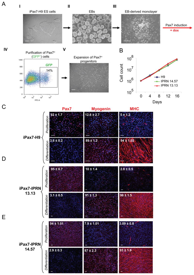
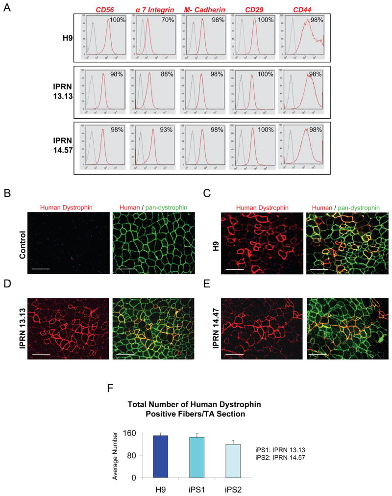
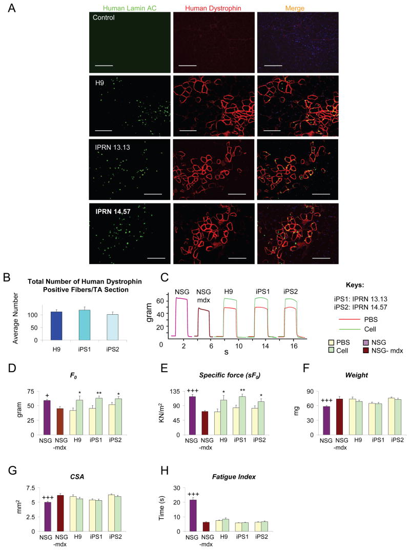
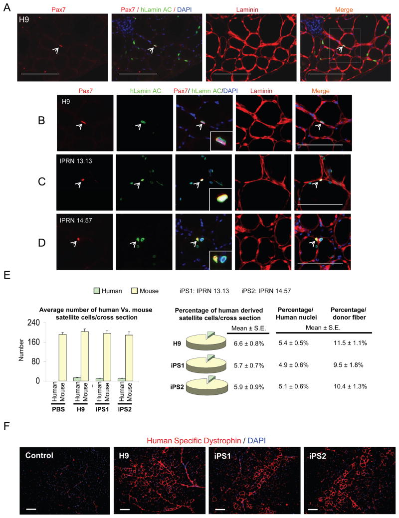
A major obstacle in the application of cell-based therapies for the treatment of neuromuscular disorders is obtaining the appropriate number of stem/progenitor cells to produce effective engraftment. The use of embryonic stem (ES) or induced pluripotent stem (iPS) cells could overcome this hurdle. However, to date, derivation of engraftable skeletal muscle precursors that can restore muscle function from human pluripotent cells has not been achieved. Here we applied conditional expression of PAX7 in human ES/iPS cells to successfully derive large quantities of myogenic precursors, which, upon transplantation into dystrophic muscle, are able to engraft efficiently, producing abundant human-derived DYSTROPHIN-positive myofibers that exhibit superior strength. Importantly, transplanted cells also seed the muscle satellite cell compartment, and engraftment is present over 11 months posttransplant. This study provides the proof of principle for the derivation of functional skeletal myogenic progenitors from human ES/iPS cells and highlights their potential for future therapeutic application in muscular dystrophies.


Figures

Figure 1
Figure 1. Myogenic induction of human ES/iPS cells by Pax7
(A) Schematic of differentiation protocol with representative morphological aspects of iPax7 H9: in the undifferentiated state as ES cell colonies in mTeSR medium (I), and in the EB stage (II). At D7 of differentiation, EBs are collected and plated on a gelatinized flask to grow as a monolayer (III). Pax7 induction is initiated at D10 of differentiation by adding dox to the myogenic medium. GFP+ (Pax7+) cells emerge in these cultures and begin to proliferate. GFP+ cells are purified by FACS (IV). Representative FACS profile shows Pax7 (GFP) expression after 4 days of dox induction in H9 differentiating ES cells. The percentage indicated represents the fraction of GFP+ cells. (IV). Pax7+ myogenic progenitors are expanded in myogenic induction medium supplemented with dox and human bFGF (V). Scale bars, 100 μm. (B) Growth curve of Pax7-induced ES- and iPS-derived myogenic progenitors during in vitro expansion. Data represent Mean ± S.E. of four independent experiments. (C–E) Immunostaining of Pax7-induced human ES- (C) and iPS- (D–E) derived myogenic cells for Pax7, Myogenin and Myosin Heavy Chain (MHC) in proliferation (upper) and differentiation (lower) conditions. With Pax7 induction under proliferation conditions, most cells express Pax7 and only a few express markers of terminal differentiation (upper panels), while under differentiation conditions (and dox withdrawal), almost all the cells become positive for Myogenin and MHC, forming multinucleated myotubes (lower panels). Cells were co-stained with DAPI (blue). Numbers on each panel represent the percentage of cells expressing Pax7, Myogenin, or MHC. Data are mean ± SE. For each condition, 4 slides were used for quantification. Scale bars, 100 μm. See also Figure S1.
Figure 2
Figure 2. Phenotypic profile and regenerative potential of human ES/iPS-derived myogenic cells
(A) Representative FACS profile of Pax7-induced human ES- and iPS-derived proliferating myogenic progenitors. Histogram plots show isotype control staining profile (gray line) versus specific antibody staining profile (red line). Percentages represent the fraction of cells that express a given surface antigen. See also Figure S2. (B–F) Transplantation of myogenic progenitors into cardiotoxin-injured NSG mice. (B) PBS-injected control muscles show no staining for human-specific dystrophin (right, in red), but as expected, uniform expression of mouse dystrophin (left, in green) as evidenced by the use of a pan-dystrophin antibody. (C–E) Engraftment of proliferating myogenic progenitors obtained from Pax7-induced human ES- (C) and iPS-derived (D–E) cells in TA muscles of NSG (n = 4 for each cell line) two months after intramuscular transplantation. Immunofluorescence staining with anti-human (in red) and anti-pan dystrophin (in green) antibodies reveals presence of donor-derived myofibers expressing human dystrophin in recipient muscles (in red). Scale bars, 100 μm. (F) Quantification of human dystrophin positive fibers in engrafted muscles shows similar engraftment of human ES- versus iPS-derived myogenic progenitors. For this, the total number of human dystrophin positive fibers in cross-sections of TA muscles (sections spanned entire muscles) was counted. Data are shown as Mean ± SE.
Figure 3
Figure 3. Efficient engraftment and functional recovery following transplantation of human ES/iPS-derived myogenic progenitorsinto dystrophic mice
(A) No staining for human Lamin AC or dystrophin is detected in PBS-injected control TA muscles of NSG-mdx mice. Cell engraftment in NSG −mdx4Cv mice (n=5 for H9, n=6 for IPRN13.13 and n=7 for IPRN 14.47). Abundant expression for human Lamin AC (in green) and dystrophin (in red) is observed in dystrophic muscles treated with human ES/iPS-derived myogenic progenitors one month after the transplantation. Note that nuclear Lamin AC staining occurs predominantly within human dystrophin+ myofibers. Scale bars, 100 μm. See also Figure S3. (B) Quantification of human dystrophin positive fibers in NSG-mdx4Cv engrafted mice shows comparable engraftment of human ES- versus iPS-derived myogenic progenitors. For this, the total number of human dystrophin positive fibers in cross-sections of TA muscles (sections spanned entire muscles) was counted. Data are shown as Mean ± SE. (C) Representative example of force tracings in TA muscles of non-treated non-injured NSG (purple line) and NSG-mdx4Cv (brown line) mice as well as CTX-injured NSG-mdx4Cv mice that had been injected with PBS (control, red line) or human ES/iPS-derived myogenic progenitors (green line). (D–E) Effect of iPax7 human ES/iPS-derived myogenic cell transplantation on absolute and specific (sF0: F0 normalized to CSA) force, respectively. Values for non-treated, non-injured NSG (purple) and NSG-mdx4Cv (brown) mice are shown for reference. (F–G) Weight and CSA of control and transplanted muscles, respectively. Values for non-treated, non-injured NSG (purple) and NSG-mdx4Cv (brown) mice are shown for reference. See also Figure S4. (H) Fatigue index: time for force to decline to 30% of its maximal value shows no significant recovery with cell treatment compared to PBS control. * P<0.05, ** P<0.01 compared to its PBS control +P<0.05, +++ P<0.001 compared to NSG-mdx4Cv
Figure 4
Figure 4. Satellite cell engraftment by human ES/iPS-derived myogenic cells
(A–B) Representative images show staining for satellite cells in muscle sections from NSG-mdx4Cv mice that had been transplanted with H9 human ES-derived myogenic progenitors. Images are shown at lower (A) and higher (B) magnification. Immunostaining shows the presence of human Lamin AC+ (in green) cells in engrafted regions. Arrows shows the presence of human-derived satellite cells in engrafted muscles, as evidenced by the presence of Pax7+ (in red) Lamin AC+ (in green) cells under the basal lamina. Scale bars, 100 μm. (C–D) Similar satellite cell engraftment was observed upon transplantation of human iPS-derived myogenic progenitors, IPRN 13.13 (C) and IPRN 14.57 (D). Scale bars, 100 μm. (E) Quantification of Pax7+Lamin AC+ and Pax7+Lamin AC cells in transplanted muscles, representative of donor human derived and host satellite cells, respectively. The total number of Pax7+Lamin AC+ and Pax7+Lamin AC cells in cross-sections of TA muscles was counted. Sections spanned entire muscles. Data are shown as Mean ± SE. Left panels show absolute numbers and right panels indicate respective percentages. Data are also shown as percentage per human nuclei as well as percentage per donor fiber. (F) Assessment of long-term engraftment at 46 weeks after transplantation in NSG mice. Immunofluorescence staining with anti-human dystrophin antibody reveals the presence of donor-derived myofibers expressing human dystrophin (in red) in NSG recipient muscles. Scale bars, 100 μm. See also Figure S4.

References

    1. Alipio Z, Liao W, Roemer EJ, Waner M, Fink LM, Ward DC, Ma Y. Reversal of hyperglycemia in diabetic mouse models using induced-pluripotent stem (iPS)-derived pancreatic beta-like cells. Proc Natl Acad Sci U S A. 2010;107:13426–13431. - PMC - PubMed
    1. Barberi T, Bradbury M, Dincer Z, Panagiotakos G, Socci ND, Studer L. Derivation of engraftable skeletal myoblasts from human embryonic stem cells. Nat Med. 2007;13:642–648. - PubMed
    1. Bosnakovski D, Xu Z, Gang EJ, Galindo CL, Liu M, Simsek T, Garner HR, Agha-Mohammadi S, Tassin A, Coppee F, et al. An isogenetic myoblast expression screen identifies DUX4-mediated FSHD-associated molecular pathologies. EMBO J. 2008;27:2766–2779. - PMC - PubMed
    1. Cornelison DD, Wold BJ. Single-cell analysis of regulatory gene expression in quiescent and activated mouse skeletal muscle satellite cells. Dev Biol. 1997;191:270–283. - PubMed
    1. Coulton GR, Rogers B, Strutt P, Skynner MJ, Watt DJ. In situ localisation of single-stranded DNA breaks in nuclei of a subpopulation of cells within regenerating skeletal muscle of the dystrophic mdx mouse. J Cell Sci. 1992;102(Pt 3):653–662. - PubMed

Publication types