. 2012 May 2;15(5):764-77.
doi: 10.1016/j.cmet.2012.04.005.

Muscle-specific deletion of carnitine acetyltransferase compromises glucose tolerance and metabolic flexibility

Affiliations

Muscle-specific deletion of carnitine acetyltransferase compromises glucose tolerance and metabolic flexibility

Deborah M Muoio et al. Cell Metab. .

Abstract

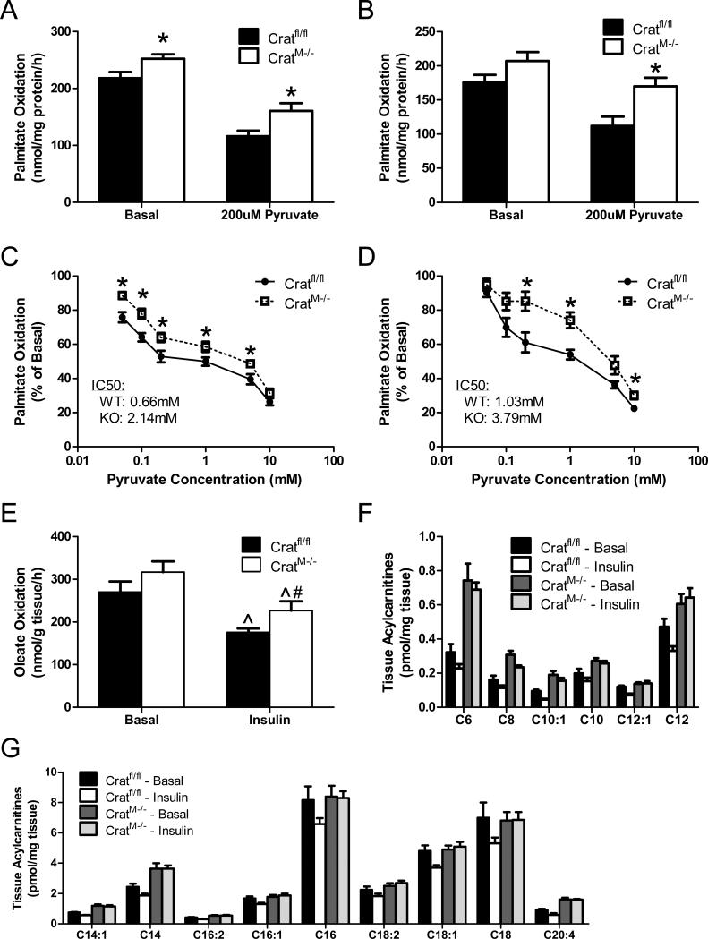
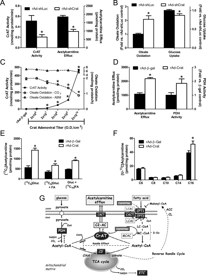
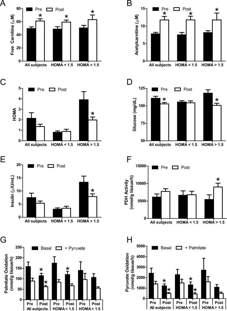
The concept of "metabolic inflexibility" was first introduced to describe the failure of insulin-resistant human subjects to appropriately adjust mitochondrial fuel selection in response to nutritional cues. This phenomenon has since gained increasing recognition as a core component of the metabolic syndrome, but the underlying mechanisms have remained elusive. Here, we identify an essential role for the mitochondrial matrix enzyme, carnitine acetyltransferase (CrAT), in regulating substrate switching and glucose tolerance. By converting acetyl-CoA to its membrane permeant acetylcarnitine ester, CrAT regulates mitochondrial and intracellular carbon trafficking. Studies in muscle-specific Crat knockout mice, primary human skeletal myocytes, and human subjects undergoing L-carnitine supplementation support a model wherein CrAT combats nutrient stress, promotes metabolic flexibility, and enhances insulin action by permitting mitochondrial efflux of excess acetyl moieties that otherwise inhibit key regulatory enzymes such as pyruvate dehydrogenase. These findings offer therapeutically relevant insights into the molecular basis of metabolic inflexibility.


Figures

Figure 1
Figure 1. Generation of mice with muscle-specific CrAT deficiency
Panel A shows the genetic strategy for generating mice with muscle-specific ablation of CrAT (CratM-/-). Tissue-specific deletion was confirmed by analyzing CrAT mRNA (panel B), protein (panel C) and enzyme activity (panel D) in multiple tissues (n=3-6/group). Enzymatic activity in isolated mitochondrial lysates from mixed gastrocnemius skeletal muscle (SkM) was measured with several short and medium chain acyl-CoAs as substrates (panel E; n=6/group).
Figure 2
Figure 2. Muscle-specific CrAT deficiency affects whole body metabolic homeostasis
CratM-/- mice and control (Cratfl/fl) littermates were fed either a 10% fat (Low Fat) or a 45% fat (High Fat) diet beginning at 5 weeks of age. Body weight (panel A) and fat mass (panel B) were measured every two weeks (n=10-13/group). Glucose tolerance tests were performed using 40 mg glucose/mouse (panel C) or 20 mg glucose/mouse (panel D) (n=10/group). For insulin tolerance tests (panel E) blood glucose levels were measured 10 min after injection of 0.04U insulin/mouse (low fat diet) or 0.08U/mouse (high fat diet) in fed mice (n=10/group). The following measures were from mice fed a low fat diet. Plasma triglycerides (TAG) and non-esterified fatty acids (NEFA) were measured following a 4 h fast (panel F; n=12/group). Following a 7 day acclimatization period, respiratory exchange ratio (RER) was monitored by indirect calorimetry immediately after an insulin injection of 0.04U/mouse (panel G). Insulin signaling (Akt total, pThr308, and pSer473) was measured using lysates prepared from gastrocnemius muscles collected 10min after insulin injection (panel H). Quantified bands were normalized to a MemCode loading control and phosphorylated proteins were expressed relative to total Akt. * Genotype effect (p< 0.05) detected by Student's t-test.
Figure 3
Figure 3. Free carnitine and acylcarnitine profiling in CratM-/- mice
Tissue and blood specimens were obtained from anesthetized mice (n=6-12/group) following a 4 h fast. Free carnitine and acylcarnitine levels in plasma (panel A), gastrocnemius muscle (SkM; panel B) and heart (panel C) were measured by mass spectrometry. The acyl chain length (C) is denoted by the corresponding metabolite number (e.g. C0= free carnitine, C2= acetylcarnitine; C3= proprionylcarnitine). Data are expressed as mean ± SEM. * Genotype effect (p< 0.05) detected by Student's t-test.
Figure 4
Figure 4. CrAT regulates PDH activity
Gastrocnemius muscles were harvested after a 4 h fast and lysates were prepared for immunoblot analysis of the mitochondrial marker protein, citrate synthase (CS), and electron transport chain complexes I, II and V. (panel A). Mitochondria were isolated from gastrocnemius muscles and a portion of the suspension was used to assess citrate synthase activity (panel B; n=12). State 3 and state 4 mitochondrial respiration rates were measured using BD Oxygen Biosensor plates after addition of 10 mM pyruvate and 5 mM malate ± 1 mM carnitine (panel C; n=6). RCR values were 7.2-7.6 for all groups. State 3 respiration was also measured with 10 mM pyruvate ± 1 mM carnitine (panel D; n=6). PDH activity was determined by measuring 14CO2 produced from 1 mM [1-14C]pyruvate ± 5 mM carnitine in isolated muscle mitochondria from Cratfl/fl and CratM-/- mice (panel E; n=6). The acute effect of 5 mM carnitine on PDH activity was evaluated in mitochondria from rat gastrocnemius muscle compared to rat liver (n=3) (panel F). Soleus muscles (n=6) were incubated 2 h with 10 mM [13C6]glucose and 200 μM fatty acid ± 100 nM insulin, followed by LCMS-based mass analysis of [12C]acetylcarnitine (C2), [13C2]acetylcarnitine ([13C2]C2) and total acetylcarnitine (C2+([13C2]C2) in the muscle tissue (panel G) and incubation buffer (panel H). * Genotype and # carnitine effects (p< 0.05) detected by Student's t-test.
Figure 5
Figure 5. CrAT deficiency promotes fat oxidation
Fatty acid oxidation to CO2 was measured in isolated muscle (gastrocnemius) mitochondria exposed to moderate (100 μM; panel A) or high (300 μM; panel B) doses of [1-14C]palmitate ± 200 μM pyruvate (n=12). Dose-dependent pyruvate inhibition of fatty acid oxidation was measured during exposure to 100 μM (panel C) or 300 μM (panel D) [1-14C]palmitate. Results were normalized to basal oxidation rates and IC50 values for the inhibitory effect of pyruvate on palmitate oxidation were calculated using GraphPad Prism software. Total oxidation of 500 μM [1-14C]oleate (panel E) was measured in intact soleus muscles (n=6) incubated 2 h in a modified KHB buffer containing 5 mM glucose ± 100 nM insulin (panel E). Soleus muscles (n=6) were incubated 2 h with 10 mM [13C6]glucose and 200 μM fatty acid ± 100 nM insulin, followed by LCMS-based mass analysis of medium (panel F) and long chain (panel G) acylcarnitines. Data are expressed as mean ± SEM. Main effects of insulin (^) and genotype (#) were detected by two-way ANOVA, p< 0.05; * post hoc test for genotype. Two-way ANOVA and MANOVA revealed main effects of insulin and genotype on the medium/long chain acylcarnitine profiles; symbols were omitted for simplicity.
Figure 6
Figure 6. CrAT counter-regulates glucose and fatty acid metabolism in primary human skeletal myocytes
Treatment of primary human skeletal myotubes with Ad-shCrAT lowered CrAT activity and acetylcarnitine efflux into the culture media (panel A), increased oxidation of 100 μM [1-14C]oleate to CO2 and decreased uptake of [3H]-2-deoxy-glucose (panel B) as compared to the rAd-shLuc control. Treatment of myotubes treated with increasing doses of rat CrAT (rAd-CrAT) increased CrAT activity and lowered complete oxidation (14CO2) of 100 μM [1-14C]oleate without affecting incomplete oxidation (acid soluble metabolites; ASM). Overexpression of CrAT increased acetylcarnitine efflux into the culture medium and increased PDH activity measured in cell lysates (panel D). Cellular levels of glucose-derived acetylcarnitine was measured after 4 h exposure to [13C6]glucose/[13C3]pyruvate mixture (5 mM/0.1 mM) ± 100 μM palmitate (FA) (panel E). Fatty acid-derived acetylcarnitine (panel E) and medium/long chain acylcarnitines (panel F) were measured after 4 h exposure to [13C16]palmitate (100μM) in the presence of unlabeled glucose/pyruvate (5 mM/0.1 mM). Data are expressed as mean ± SEM and are representative of 2-4 independent experiments. * Effect of CrAT silencing or overexpression (p< 0.05) detected by Student's t-test. Proposed model of CrAT-mediated regulation of mitochondrial carbon trafficking and metabolic flexibility (panel G). During the early phases of re-feeding, muscle mitochondria are confronted with a heavy influx of both glucose and fatty acid fuel. By permitting mitochondrial efflux of excess acetyl moieties, CrAT allows PDH activity to increase despite high rates of β-oxidation. This eases the Randle effect and facilitates a rapid transition from fatty acid to glucose oxidation. CrAT deficiency and/or carnitine insufficiency imposes a more rigid version of the Randle cycle, wherein feeding-induced stimulation of PDH activity is heavily dependent on the production of malonyl-CoA and resultant inhibition of CPT1. Abbreviations: Acetyl-CoA carboxylase (ACC); acetylcarnitine (C2-AC); carnitine acylcarnitine translocase (CACT); carnitine palmitoyltransferase 1 (CPT1); carnitine palmitoyltransferase 2 (CPT2); citrate lyase (CL); carnitine acetyltransferase (CrAT); citrate synthase (CS); electron transport chain (ETC); inner mitochondrial membrane (imm); long chain acylcarnitine (LCAC); long chain acyl-CoA (LC-CoA); medium chain acyl-CoA (MC-CoA); outer mitochondrial membrance (omm); oxaloacetic acid (OAA); pyruvate dehydrogenase (PDH); tricarboxylic acid (TCA).
Figure 7
Figure 7. Carnitine supplementation improves glucose homeostasis in insulin resistant humans
Older aged human subjects (n=14) with modestly elevated fasting blood glucose levels received supplemental L-carnitine (2 g/day) for 6 months. Fasting serum samples were collected pre- and post-intervention for measurement of free carnitine (panel A), acetylcarnitine (panel B), HOMA (panel C), glucose (panel D) and insulin (panel E). Tissue homogenates prepared from vastus lateralis biopsies were used to measure PDH activity with 5 mM [1-14C]pyruvate (panel F). Substrate switching was determined by measuring oxidation of 50μM [1-14C]palmitate (panel G) or 5 mM [2-14C]pyruvate (panel H) to 14CO2 in the absence or presence of the unlabeled competing substrate (5 mM pyruvate or 100μM palmitate, respectively). Results were stratified according to HOMA at enrollment, using a cut off of 1.5; n=8 below and n=6 above. * Effect of carnitine supplementation analyzed by 1-tailed paired Student's t-test (p<0.05).

References

    1. Albarado DC, McClaine J, Stephens JM, Mynatt RL, Ye J, Bannon AW, Richards WG, Butler AA. Impaired coordination of nutrient intake and substrate oxidation in melanocortin-4 receptor knockout mice. Endocrinology. 2004;145:243–252. - PubMed
    1. An J, Muoio DM, Shiota M, Fujimoto Y, Cline GW, Shulman GI, Koves TR, Stevens R, Millington D, Newgard CB. Hepatic expression of malonyl-CoA decarboxylase reverses muscle, liver and whole-animal insulin resistance. NatMed. 2004;10:268–274. - PubMed
    1. Arumugam R, Horowitz E, Noland RC, Lu D, Fleenor D, Freemark M. Regulation of islet beta-cell pyruvate metabolism: interactions of prolactin, glucose, and dexamethasone. Endocrinology. 2010;151:3074–3083. - PMC - PubMed
    1. Becker TC, BeltrandelRio H, Noel RJ, Johnson JH, Newgard CB. Overexpression of hexokinase I in isolated islets of Langerhans via recombinant adenovirus. Enhancement of glucose metabolism and insulin secretion at basal but not stimulatory glucose levels. J Biol Chem. 1994;269:21234–21238. - PubMed
    1. Beenakkers AM, Klingenberg M. Carnitine-Coenzyme a Transacetylase in Mitochondria from Various Organs. Biochim Biophys Acta. 1964;84:205–207. - PubMed

Publication types

MeSH terms