. 2012 Jul;33(21):5414-22.
doi: 10.1016/j.biomaterials.2012.04.032. Epub 2012 May 3.

Noninvasive monitoring of orthotopic glioblastoma therapy response using RGD-conjugated iron oxide nanoparticles

Affiliations

Noninvasive monitoring of orthotopic glioblastoma therapy response using RGD-conjugated iron oxide nanoparticles

Fan Zhang et al. Biomaterials. 2012 Jul.

Abstract

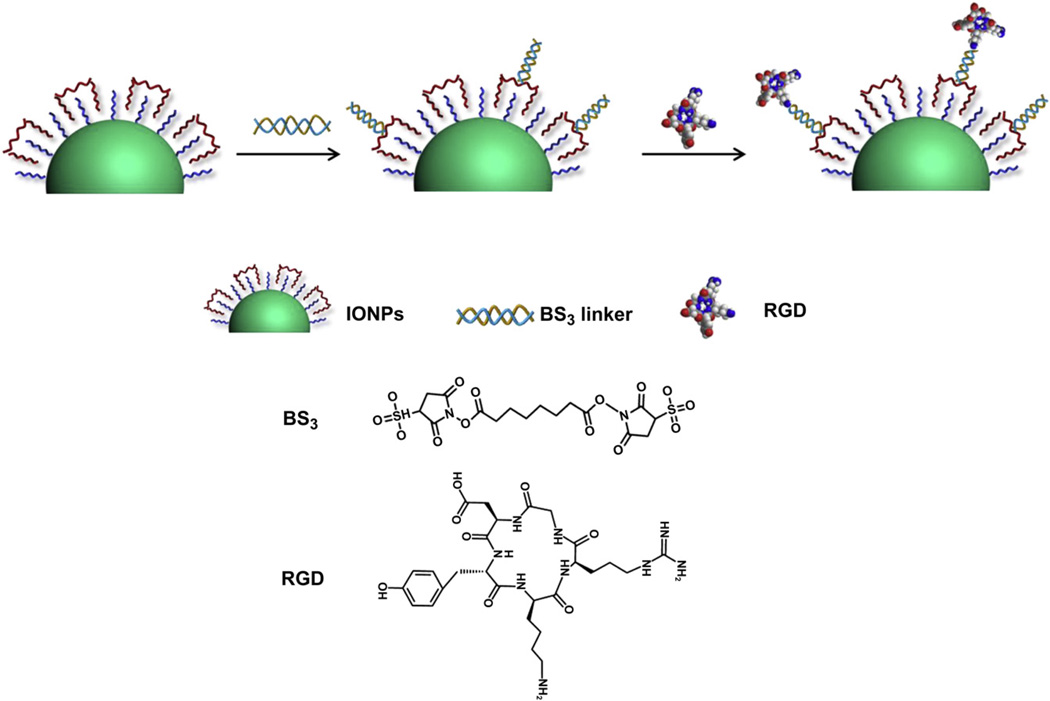
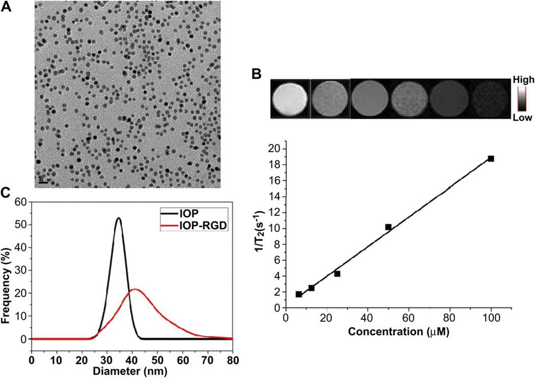
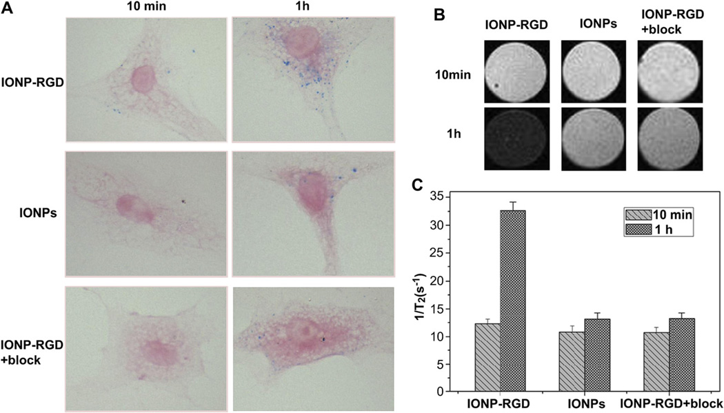
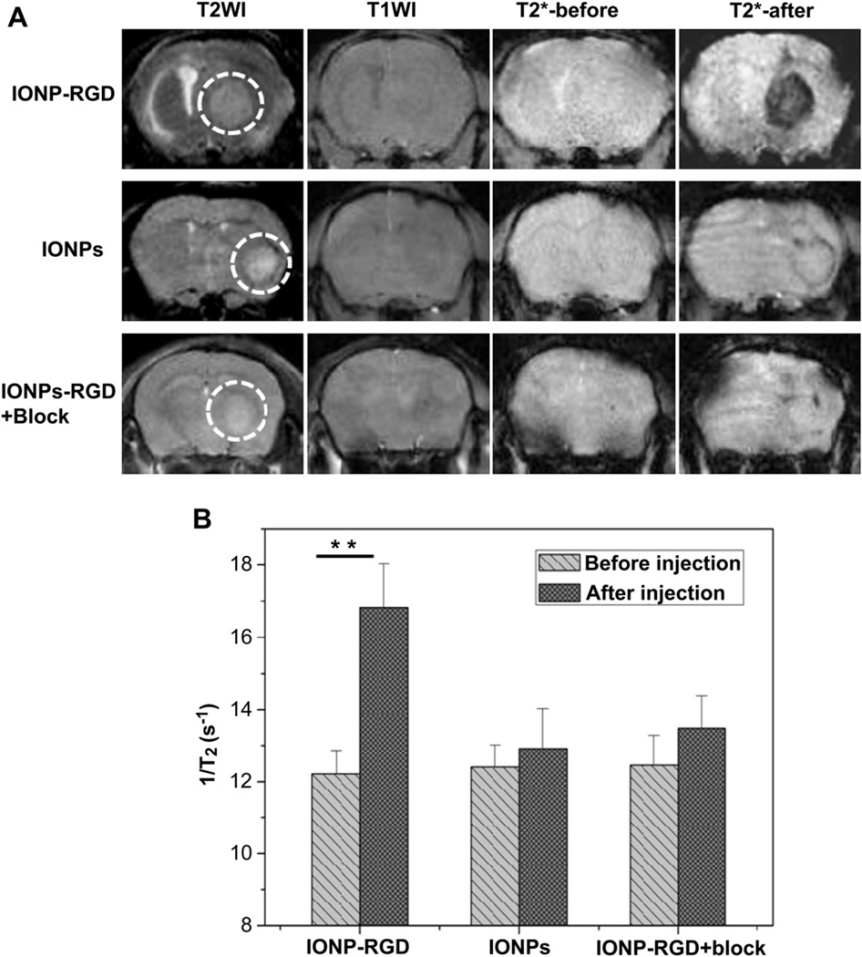
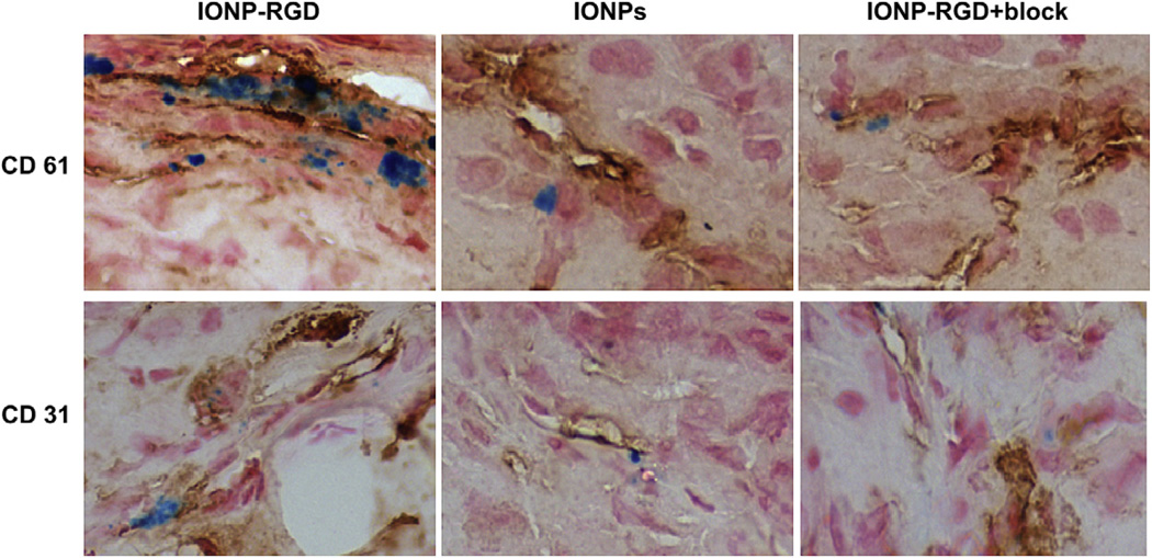
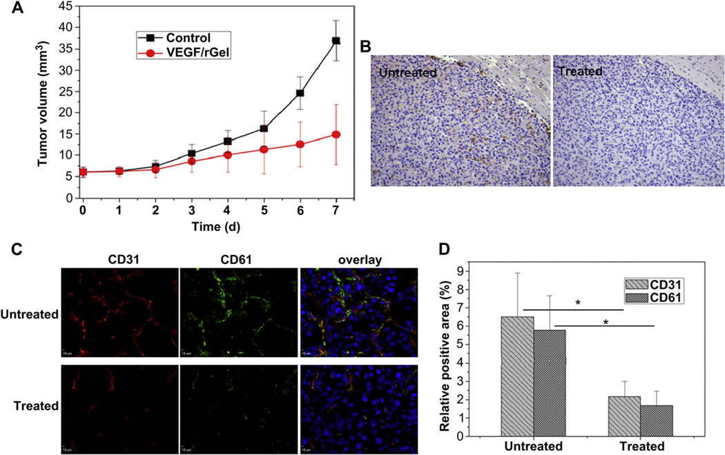
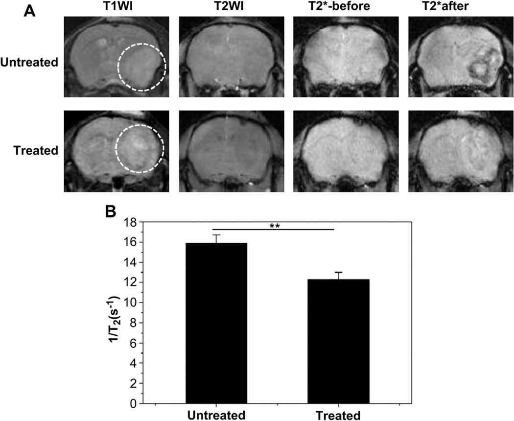
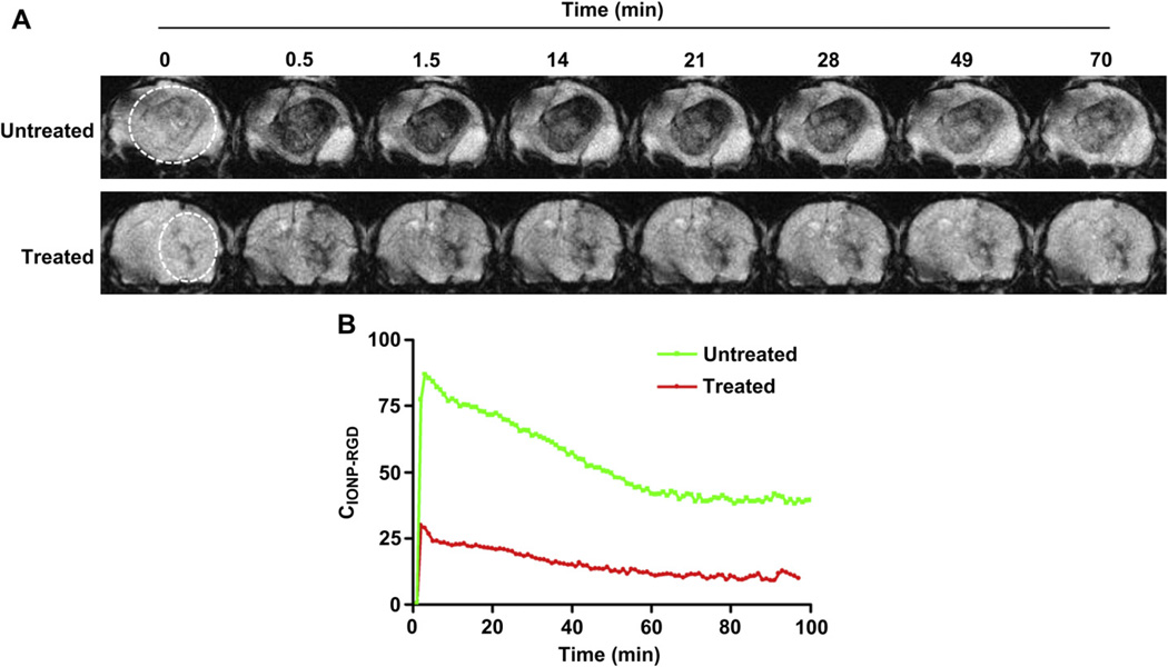
Noninvasive imaging techniques have been considered important strategies in the clinic to monitor tumor early response to therapy. In the present study, we applied RGD peptides conjugated to iron oxide nanoparticles (IONP-RGD) as contrast agents in magnetic resonance imaging (MRI) to noninvasively monitor the response of a vascular disrupting agent VEGF(121)/rGel in an orthotopic glioblastoma model. RGD peptides were firstly coupled to IONPs coated with a crosslinked PEGylated amphiphilic triblock copolymer. In vitro binding assays confirmed that cellular uptake of particles was mainly dependent on the interaction between RGD and integrin α(v)β(3) of human umbilical vein endothelial cells (HUVEC). The tumor targeting of IONP-RGD was observed in an orthotopic U87 glioblastoma model. Finally, noninvasive monitoring of the tumor response to VEGF(121)/rGel therapy at early stages of treatment was successfully accomplished using IONP-RGD as a contrast agent for MRI, a superior method over common anatomical approaches which are based on tumor size measurements. This preclinical study can accelerate anticancer drug development and promote clinical translation of nanoprobes.


Figures

Fig. 1
Fig. 1
Schematic illustration of the conjugation of IONPs with RGD peptides.
Fig. 2
Fig. 2
Characterization of IONPs and IONP-RGD (A) TEM of IONPs (B) T2*-weighted phantom images of IONPs at different Fe concentration. Top, T2*-weighted phantom images; Bottom, 1/T2 vs. Fe concentration curve of IONPs.
Fig. 3
Fig. 3
Cellular uptake of particles in HUVECs. The IONPs, IONP-RGD and IONP-RGD + block were incubated with HUVECs for 10 min and 1 h, respectively. Cellular uptake was evaluated by (A) Prussian blue staining and (B) T2-weighted phantom images (C) Quantitative analysis of relaxation time of T2*-weighted phantom images.
Fig. 4
Fig. 4
T2*-weighted MR images of nude mice bearing orthotopic U87MG glioblastoma (A) T2*-weighted MR images were acquired before and after injection of IONPs, IONP-RGD and IONP-RGD + block, respectively (B) Quantitative analysis of T2*-weighted MR images in tumor areas.
Fig. 5
Fig. 5
Prussian blue and CD31/CD61 double staining of the tumor sections. At 6 h post-injection, the mice were sacrificed and frozen tissue slices were prepared.
Fig. 6
Fig. 6
The effect of VEGF121/rGel on the integrin αvβ3 expression of tumor angiogenic blood vessels (A) Tumor growth curves of untreated and VEGF121/rGel treated groups were analyzed by MRI (B) CD31 staining of tumor angiogenic blood vessels by immnohistochemistry (C) CD31/CD61 double staining of tumor angiogenic blood vessels and integrin αvβ3 expression on frozen tissue slices (D) Quantitative analysis of CD31- and CD 61-positive area using Image J software (5 mice each group).
Fig. 7
Fig. 7
Monitoring of therapeutic response of VEGF121/rGel using IONP-RGD in orthotopic U87MG glioblastoma model (A) T2*-weighted MR images of untreated and VEGF121/rGel treated groups were acquired before and after injection of IONP-RGD (B) Quantification analysis of T2*-weighted MR images. The white circle indicates location of the implanted tumor (4 mice each group).
Fig. 8
Fig. 8
Dynamic monitoring of therapeutic response of VEGF121/rGel using IONP-RGD in orthotopic U87MG glioblastoma model (A) T2*-weighted MR dynamic images of untreated and VEGF121/rGel treated groups were acquired before and after injection of IONP-RGD (B) Quantitative analysis of T2*-weighted MR dynamic images (4 mice each group).

References

    1. Weber WA. Assessing tumor response to therapy. J Nucl Med. 2009;1(50 Suppl):1S–10S. - PubMed
    1. Husband JE, Schwartz LH, Spencer J, Ollivier L, King DM, Johnson R, et al. Evaluation of the response to treatment of solid tumours - a consensus statement of the International Cancer Imaging Society. Br J Cancer. 2004;90(12):2256–2260. - PMC - PubMed
    1. Han Z, Fu A, Wang H, Diaz R, Geng L, Onishko H, et al. Noninvasive assessment of cancer response to therapy. Nat Med. 2008;14(3):343–349. - PubMed
    1. Day SE, Kettunen MI, Gallagher FA, Hu DE, Lerche M, Wolber J, et al. Detecting tumor response to treatment using hyperpolarized 13C magnetic resonance imaging and spectroscopy. Nat Med. 2007;13(11):1382–1387. - PubMed
    1. de Langen AJ, van den Boogaart V, Lubberink M, Backes WH, Marcus JT, van Tinteren H, et al. Monitoring response to antiangiogenic therapy in non-small cell lung cancer using imaging markers derived from PET and dynamic contrast-enhanced MRI. J Nucl Med. 2011;52(1):48–55. - PubMed

Publication types

MeSH terms