. 2012 Jun 6;20(6):1007-18.
doi: 10.1016/j.str.2012.03.011. Epub 2012 May 3.

Structural basis for polyadenosine-RNA binding by Nab2 Zn fingers and its function in mRNA nuclear export

Affiliations

Structural basis for polyadenosine-RNA binding by Nab2 Zn fingers and its function in mRNA nuclear export

Christoph Brockmann et al. Structure. .

Abstract

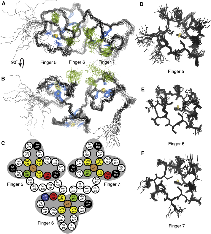
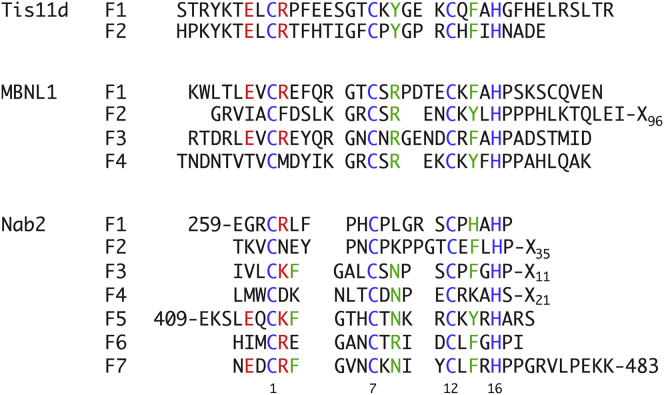
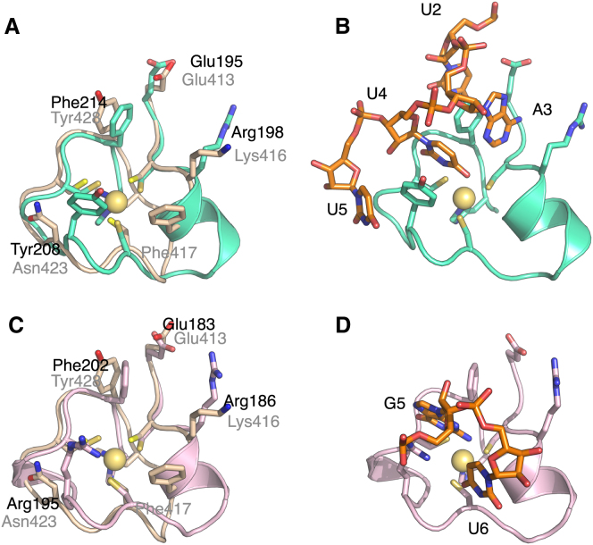
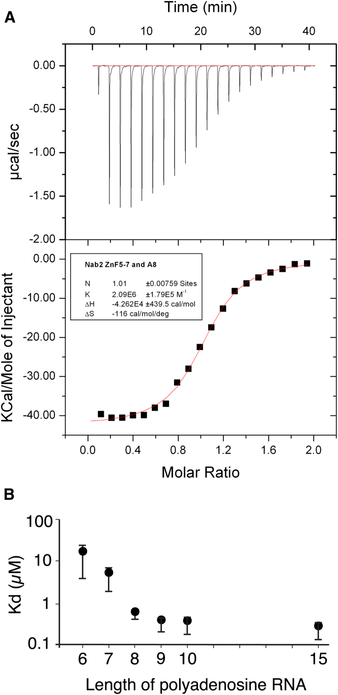
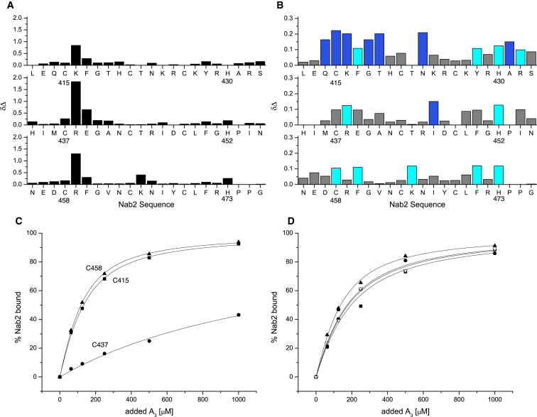
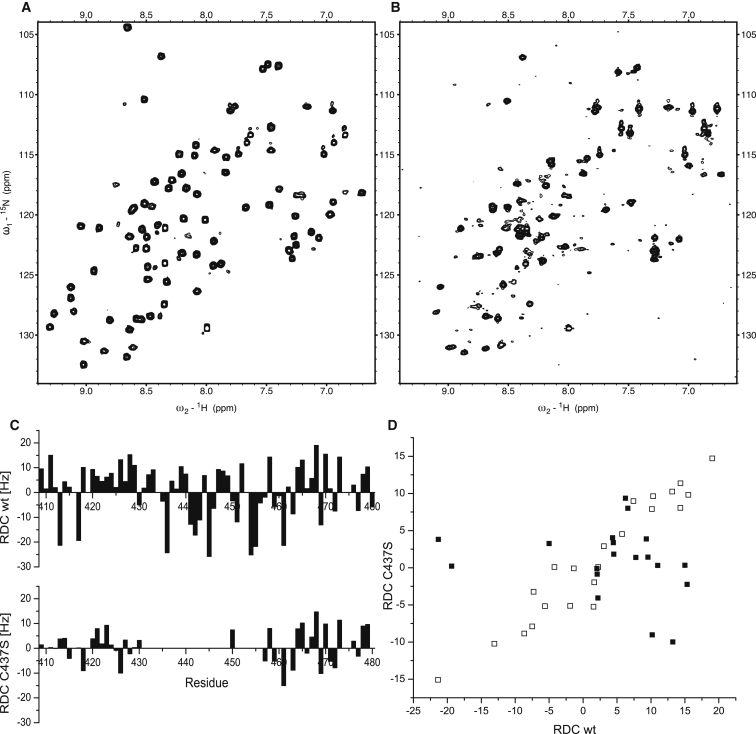
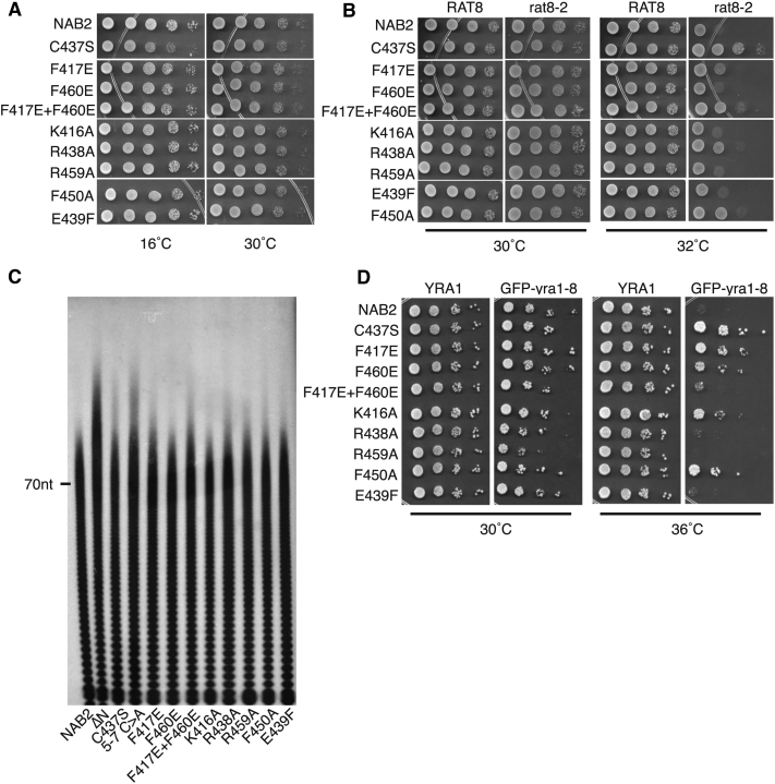
Polyadenylation regulation and efficient nuclear export of mature mRNPs both require the polyadenosine-RNA-binding protein, Nab2, which contains seven CCCH Zn fingers. We describe here the solution structure of fingers 5-7, which are necessary and sufficient for high-affinity polyadenosine-RNA binding, and identify key residues involved. These Zn fingers form a single structural unit. Structural coherence is lost in the RNA-binding compromised Nab2-C437S mutant, which also suppresses the rat8-2 allele of RNA helicase Dbp5. Structure-guided Nab2 variants indicate that dbp5(rat8-2) suppression is more closely linked to hyperadenylation and suppression of mutant alleles of the nuclear RNA export adaptor, Yra1, than to affinity for polyadenosine-RNA. These results indicate that, in addition to modulating polyA tail length, Nab2 has an unanticipated function associated with generating export-competent mRNPs, and that changes within fingers 5-7 lead to suboptimal assembly of mRNP export complexes that are more easily disassembled by Dbp5 upon reaching the cytoplasm.


Figures

Figure 1
Figure 1
NMR Structure of Nab2 ZnF5-7 (A and B) Ensembles of the NMR structure of Nab2 residues 409–483. Of 50 calculated structures, 20 were selected by lowest total energy. The structures are superimposed based on the backbone of residues 410–480. Side chains of the Zn-coordinating residues are shown in blue. The RNA-binding residues on ZnFs 5 and 7 (see text) are shown in green. The three fingers associate to form a novel single structural domain in which individual fingers pack together to produce a pseudo-helical arrangement. (C) Schematic illustration of the structure of fingers 5-7. Three cysteines (yellow) and a histidine (green) bind the Zn ion (brown) in each finger. Isoleucines 435 and 454, located in the linkers between fingers, are buried in the interfaces between the fingers. Residues located in finger 5 (red, Lys 416; black, Phe417, Asn423, Tyr428) and finger 7 (red, Arg459; black, Phe460, Asn466, Phe471) show large chemical shift changes on addition of A3. Finger 6 has Glu439 (blue) instead of an aromatic residue in position 2. Mutation of Lys416 or Phe417 in finger 5, Cys437, Arg438, or Phe450 (black) in finger 6, or Arg459 or Phe460 in finger 7 decreases the affinity for polyadenosine (see Table 2). (D–F) The same ensemble of structures is shown as in (A) and (B) but is now shown superimposed on the Cα positions from the individual fingers: (D) shows ZnF5 containing residues 414–431, (E) illustrates ZnF6 containing residues 436–453, and (F) shows ZnF7 containing residues 457–474). See also Figure S1 and Movie S1.
Figure 2
Figure 2
Alignment of the Sequences of Related ZnFs Shown are the seven ZnFs of Nab2 itself, as well as the closely related fingers from the proteins Tis11d and MBNL1. Residue positions within each finger are numbered taking the first cysteine residue as position 1. See also Figure S2.
Figure 3
Figure 3
Comparison of Nab2 with Other Tandem ZnF Domains (A) Structural alignment of Nab2 ZnF5 (wheat) with ZnF2 of the Tis11d tandem ZnF domain (shown in cyan) (PDB 1RGO). Side-chain residues involved in RNA binding in the Tis11d structure are shown. (B) RNA (orange)-bound structure of Tis11d. (C) Structural alignment of Nab2 ZnF5 (shown as wheat) with ZnF3 of the MBNL1 tandem ZnF domain (shown in pink) (PDB 3D2S). Side-chain residues involved in RNA binding in chain (A) in the MBNL1 are shown. (D) RNA (orange)-bound structure of MBNL1.
Figure 4
Figure 4
Affinity of Polyadenosine-RNA for Nab2 ZnF5-7 (A) ITC raw data (upper panel) for binding of A8 to Nab2 ZnF5-7 with integrated peaks and fitting curve (lower panel). (B) Influence of the length (A6–A10) of polyadenosine-RNA on the affinity for Nab2 ZnF5-7. Error bars represent SD from three independent measurements.
Figure 5
Figure 5
Binding of Nab2 ZnF5-7 to Polyadenosine-RNA (A) CSP of backbone amide groups of wild-type ZnF5-7 in the presence of 5 mM AMP. (B) CSP of backbone amide groups of wild-type Nab2 fingers 5-7 in the presence of 250 μM A3. (C) Normalized binding isotherms extracted from the titration data summarized in (B), following residues C415 (■), C437 (●), and C458 (▴). (D) Binding isotherms extracted from the titration of the E439F mutant with A3, following residues C415 (■), H420 (▿), C437 (●), and C458 (▴).
Figure 6
Figure 6
Effects of the C437S Mutant on the Structure of Nab2 ZnF5-7 (A) 15N-HSQC spectrum of wild-type Nab2409–483. (B) The same spectrum as shown in (A) but for the C437S Nab2 mutant. Comparison of (A) and (B) showed that peaks close to the original positions of the finger 5 and 7 amides are retained, consistent with these fingers still being folded, whereas most of the peaks from ZnF6 are significantly broadened, probably indicating exchange between different conformations. (C) Comparison of backbone 1H-15N RDC values obtained for the wild-type and C437S Nab2 proteins. The absence of measurable values for ZnF6 reflects the broad or unresolved nature of the corresponding peaks in these cases, whereas the reduced magnitudes of the RDCs in ZnF7 and (especially) ZnF5 indicate a higher degree of relative mobility for these fingers in the mutant. (D) Correlation plot of N-H RDC values obtained for the wild-type and C437S Nab2 proteins. RDC data obtained for ZnF5 (residues 410–430) are shown using filled squares (■), whereas RDC data obtained for ZnF7 (residues 458–479) are shown using open squares (□). A strong correlation is visible between the RDCs measured for the wild-type and the C437S mutant for ZnF7, but such a correlation is absent for the corresponding RDC data for ZnF5. This suggests that the alignment is dominated by effects involving ZnF7 in both the wild-type and mutant proteins but that in the mutant, ZnF5 is mobile relative to ZnF7, thereby rendering the alignment for ZnF5 in the mutant independent of that for ZnF7.
Figure 7
Figure 7
Functional Analysis of Nab2 Variants In Vivo The function of Nab2 variants was assessed using a plasmid-shuffle assays such that each variant was examined as the only functional copy of the essential Nab2 protein. Results of these experiments are summarized in Table 2. For growth assays, yeast cells expressing each Nab2 variant were serially diluted and spotted on plates. (A) Nab2 ZnF variants can function in place of Nab2. Each variant supports normal cell growth at 30°C, but nab2-C437S shows slow growth at 16°C, indicating a cold-sensitive growth phenotype. (B) Suppression of the temperature-sensitive growth phenotype of dbp5(rat8-2) mutant cells by Nab2 variants. A plasmid-shuffle assay in ΔNAB2 rat8-2 mutant cells was employed to examine suppression of the temperature-sensitive growth of dbp5(rat8-2) mutant cells at 32°C. As controls, no suppression is observed with wild-type NAB2, whereas nab2-C437S suppresses robustly. (C) Bulk poly(A) tail length was examined by an RNaseA/T1 assay. Cells expressing each Nab2 variant as the sole copy of Nab2 were grown to log phase, and poly(A) tails were labeled and resolved by gel electrophoresis. The position of a 70-nucleotide (70nt) marker is indicated. (D) Suppression of the temperature-sensitive growth phenotype of GFP-yra1-8 mutant cells. A plasmid-shuffle assay in ΔNAB2 GFP-yra1-8 mutant cells was employed to examine suppression. As a control, no suppression is observed with wild-type NAB2. In contrast, nab2-C437S robustly suppresses, and some but not all of the variants suppress the temperature-sensitive growth of GFP-yra1-8 cells at 36°C. See also Figure S3.

Comment in

References

    1. Adams A., Gottschling D.E., Kaiser C.A., Stearns T. Cold Spring Harbor Laboratory Press; Cold Spring Harbor, NY: 1997. Methods in Yeast Genetics.
    1. Aitchison J.D., Blobel G., Rout M.P. Kap104p: a karyopherin involved in the nuclear transport of messenger RNA binding proteins. Science. 1996;274:624–627. - PubMed
    1. Alcázar-Román A.R., Tran E.J., Guo S., Wente S.R. Inositol hexakisphosphate and Gle1 activate the DEAD-box protein Dbp5 for nuclear mRNA export. Nat. Cell Biol. 2006;8:711–716. - PubMed
    1. Anderson J.T., Wilson S.M., Datar K.V., Swanson M.S. NAB2: a yeast nuclear polyadenylated RNA-binding protein essential for cell viability. Mol. Cell. Biol. 1993;13:2730–2741. - PMC - PubMed
    1. Baker K.E., Coller J., Parker R. The yeast Apq12 protein affects nucleocytoplasmic mRNA transport. RNA. 2004;10:1352–1358. - PMC - PubMed

Publication types

MeSH terms