. 2012 Dec;39(4):294-303.
doi: 10.1016/j.jaut.2012.03.003. Epub 2012 May 3.

Porphyromonas gingivalis promotes Th17 inducing pathways in chronic periodontitis

Affiliations

Porphyromonas gingivalis promotes Th17 inducing pathways in chronic periodontitis

Niki M Moutsopoulos et al. J Autoimmun. 2012 Dec.

Abstract

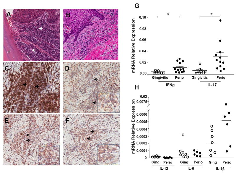
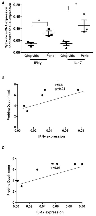
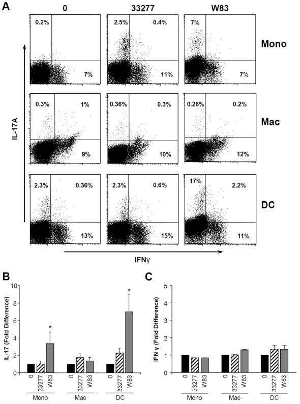
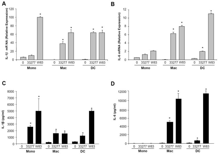
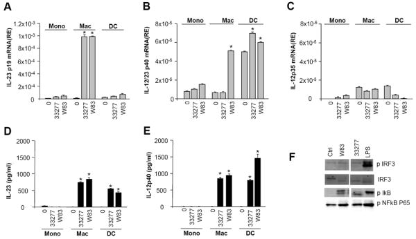
In periodontitis, a common chronic inflammatory condition, gram-negative-rich bacterial biofilms trigger, in susceptible individuals, perpetuating inflammation that results in extensive tissue damage of tooth supporting structures. To delineate immune cell-dependent mechanisms whereby bacterial challenge drives persistent destructive inflammation in periodontitis and other inflammatory diseases, we studied involved tissues ex vivo and investigated host cell responses to the periodontal pathogen Porphyromonas gingivalis, in vitro. Diseased lesions were populated by abundant Th17 cells, linked to infection, chronic inflammation/autoimmunity and tissue pathology. In vitro, P. gingivalis, particularly the more virulent strain W83, stimulated myeloid antigen presenting cells (APC) to drive Th17 polarization. Supernatants from myeloid APC exposed to P. gingivalis were capable of enhancing Th17 but not Th1 polarization. P. gingivalis favored the generation of Th17 responses by stimulating the production of Th17 related cytokines IL-1β, IL-6 and IL-23, but not Th1 related IL-12. By inducing NFκB activation, P. gingivalis promoted IL-1β, IL-6 and IL-12p40 production, but not IRF3 phosphorylation, connected to generation of the IL-12p35 chain, ultimately restricting formation of the intact IL-12 molecule. Promotion of Th17 lineage responses was also aided by P. gingivalis proteases, which appeared to differentially degrade pivotal cytokines. In this regard, IL-12 was largely degraded by P. gingivalis, whereas IL-1β was more resistant to proteolysis. Our data unveil multiple pathways by which P. gingivalis may orchestrate chronic inflammation, providing insights into interventional strategies.


Conflict of interest statement

Disclosure

The authors have no conflicts of interest.

Figures

Figure 1
Figure 1. Inflammatory infiltrate in lesions of periodontal disease
A. H&E staining reveals the dense inflammatory infiltrate (indicated by arrowheads) in areas adjacent to the tooth structure (T) in representative tissue from patients with severe periodontitis (CAL>6mm) as compared to minimally inflamed tissues from gingivitis individuals (B). C. Immunohistochemical staining using a monoclonal antibody to CD3 indicates large numbers of CD3 positive T cells (brown) in the periodontitis lesion. D-F. Immunohistochemical detection of cytokines using cytokine-specific monoclonal antibodies. Within the lesion, IFN-γ+ (D), IL-17+ (E), and IL-23+ (F) cell populations, are identified by brown staining, and indicated by arrows. All images are at original magnification 20x with inserts at magnification 40x. G. mRNA expression relative to housekeeping gene HPRT (2ˆ-ΔCT) in periodontitis (Perio, severe tissue destruction CAL>6mm, n=12) and gingivitis tissues (inflamed tissues with minimal tissue destruction, n=8) for IFN-γ and IL-17. Median ± SE indicated. *indicates significance at p<0.05, median values indicated. H. Relative mRNA expression (2ˆ-ΔCT) for IL-12, IL-6 and IL-1β in gingivitis (n=7) and periodontitis (n=6) patient tissues.
Figure 2
Figure 2. Cytokine expression in periodontitis tissues
A. mRNA expression for IFN-γ and IL-17 relative to the expression of the T cell marker CD3 is shown in gingivitis tissues (inflamed tissues with minimal tissue destruction, n=3) and in periodontitis (Perio, severe tissue destruction, n=3). Median ± SE indicated. *indicates significance at p<0.05. B-C. Correlation between tissue destruction in each sample (measured in mm, Probing Depth measure) and IFN-γ and IL-17 cytokine expression. P values and Pearson r shown for each comparison.
Figure 3
Figure 3. Th cell lineage differentiation after exposure of PBMC to supernatants from APC treated with P. gingivalis
Antigen presenting cells (APC), including donor-matched monocyte, macrophage and dendritic cells were untreated or exposed to P. gingivalis strains 33277 and W83 (1:50) for 0-24h. 24h APC supernatants (media:sup ratio 1:1) were added to PBMC which were cultured for 3-5 days in the presence of anti-CD3/anti-CD28 (3μg/ml). At the end of this period, PBMC were stimulated with PMA and ionomycin for 5 hr, in the presence of Golgistop, and cells were stained for extracellular CD3 and intracellular IFNγ and IL-17 and analyzed by flow cytometry. A. Representative dot blot showing percent IFNγ+, IL-17+, IFNγ +/IL-17+ within the CD3+ population, stimulated with aCD3/CD28 in the presence of indicated APC supernatants. B. Fold difference in percent CD3+/IL-17+ in PBMC treated with supernatants from untreated APC versus P. gingivalis exposed APC (mean±SE, n=3 experiments. C. Fold difference in percent CD3+/IFN-γ+ + in PBMC treated with supernatants from untreated APC versus P. gingivalis exposed APC (mean±SE, n=3 experiments). *indicates significance at p<0.05.
Figure 4
Figure 4. Th17 polarizing cytokines produced by APC exposed to P. gingivalis
Donor matched monocyte (mono), macrophage (mac) and dendritic cells (DC) (3×10⁶/ml) were exposed to P. gingivalis ATCC 33277 or W83 (1:50). At 1h, cells were processed for total RNA and in parallel cultures at 24h, supernatants were evaluated for cytokine levels by multiplex ELISA. A. IL-1β mRNA relative expression levels (2ˆ-ΔCT) measured by real-time PCR. B. IL-6 mRNA relative expression (2ˆ-ΔCT). C. IL-1β protein levels in 24hr culture supernatant. D. IL-6 protein levels in 24hr culture supernatant. All data representative of n=3 experiments. Significance indicated between untreated (0) and P. gingivalis exposed, * = p<0.05.
Figure 5
Figure 5. IL-23 vs IL-12 production by APC exposed to P. gingivalis
Donor matched monocyte (mono), macrophage (mac) and dendritic cells (DC) (3×10⁶/ml) were untreated or exposed to P. gingivalis ATCC 33277 or W83 at a ratio of 1:50. At 1h cells were processed for total RNA and supernatants from 24h cultures were collected and tested for cytokine levels by multiplex ELISA. A. mRNA levels (RE= relative expression) for p19 measured by real-time PCR. B. IL-12p40 mRNA expression levels. C. IL-12p35 mRNA expression levels. D. IL-23 protein levels. E. IL-12p40 protein levels. All data representative of n=3 experiments,* p<0.05, * representing significance between untreated and P. gingivalis exposed. At 30min cells were processed for protein (signaling); F. Western blots for pIRF3, IRF, pIκB, pNFκB.
Figure 6
Figure 6. Cytokine degradation by P. gingivalis
Antigen presenting cells (APC), including donor-matched monocyte, macrophage and dendritic cells were untreated or exposed to P. gingivalis strains 33277 and W83 (1:50) in the presence/absence of P. gingivalis specific protease inhibitors (gingipains KYT-1 and KYT-36, at a concentration of 106M) for 24h and supernatants were tested for concentrations of IL-1β, Il-6, IL-23 and IL-12p70. A. Supernatant cytokine levels in 24h cultures of DC exposed to P. gingivalis (2 strains) +/- PI (protease inhibitors), representative experiment shown, *indicates significance at p<0.05. B. Recombinant cytokines (IL-1β, IL-6, IL-12p70, IL-23 at 5000pg/ml) were exposed to P. gingivalis at 105-107 bacteria/ml for 10-20min. Detected cytokine levels are shown at 20 min after P.gingivalis exposure at 105 bacteria/ml. Cytokine levels expressed as a percentage of input levels. C. Protein gel of IL-1β post P.gingivalis (2 strains) exposure at 20 min. D. Protein gel of IL-12 post P. gingivalis exposure at 20 min.

References

    1. Darveau RP. Periodontitis: a polymicrobial disruption of host homeostasis. Nat Rev Microbiol. 8:481–90. - PubMed
    1. Hajishengallis G. Porphyromonas gingivalis-host interactions: open war or intelligent guerilla tactics? Microbes Infect. 2009;11:637–45. - PMC - PubMed
    1. Pussinen PJ, Alfthan G, Jousilahti P, Paju S, Tuomilehto J. Systemic exposure to Porphyromonas gingivalis predicts incident stroke. Atherosclerosis. 2007;193:222–8. - PubMed
    1. Moen K, Brun JG, Madland TM, Tynning T, Jonsson R. Immunoglobulin G and A antibody responses to Bacteroides forsythus and Prevotella intermedia in sera and synovial fluids of arthritis patients. Clin Diagn Lab Immunol. 2003;10:1043–50. - PMC - PubMed
    1. Martinez-Martinez RE, Abud-Mendoza C, Patino-Marin N, Rizo-Rodriguez JC, Little JW, Loyola-Rodriguez JP. Detection of periodontal bacterial DNA in serum and synovial fluid in refractory rheumatoid arthritis patients. J Clin Periodontol. 2009;36:1004–10. - PubMed

Publication types

MeSH terms