. 2012 Jul;1821(7):1012-21.
doi: 10.1016/j.bbalip.2012.04.006. Epub 2012 Apr 27.

The structure of monoacylglycerol lipase from Bacillus sp. H257 reveals unexpected conservation of the cap architecture between bacterial and human enzymes

Affiliations

The structure of monoacylglycerol lipase from Bacillus sp. H257 reveals unexpected conservation of the cap architecture between bacterial and human enzymes

Srinivasan Rengachari et al. Biochim Biophys Acta. 2012 Jul.

Abstract

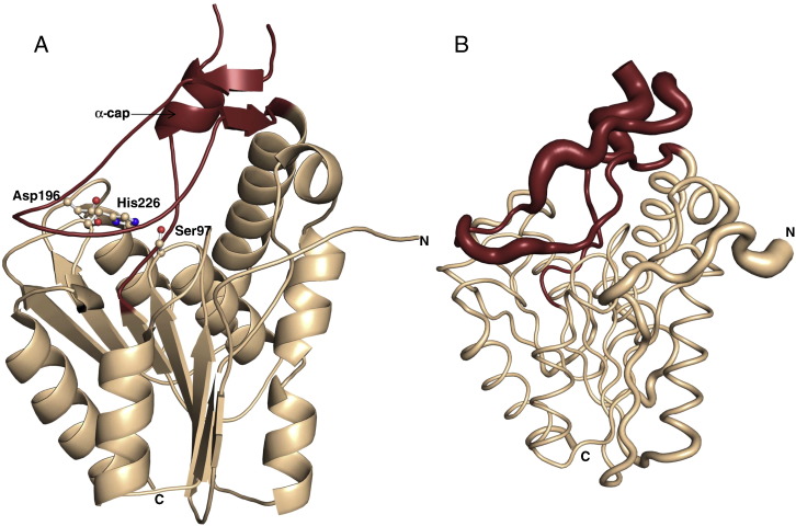
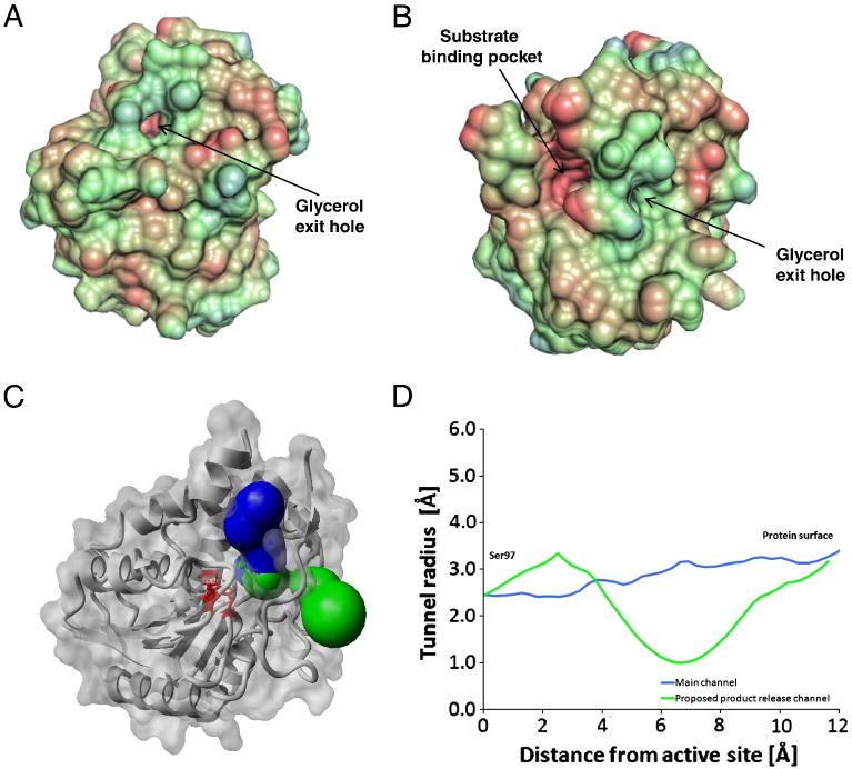
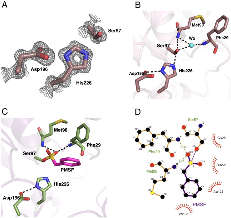
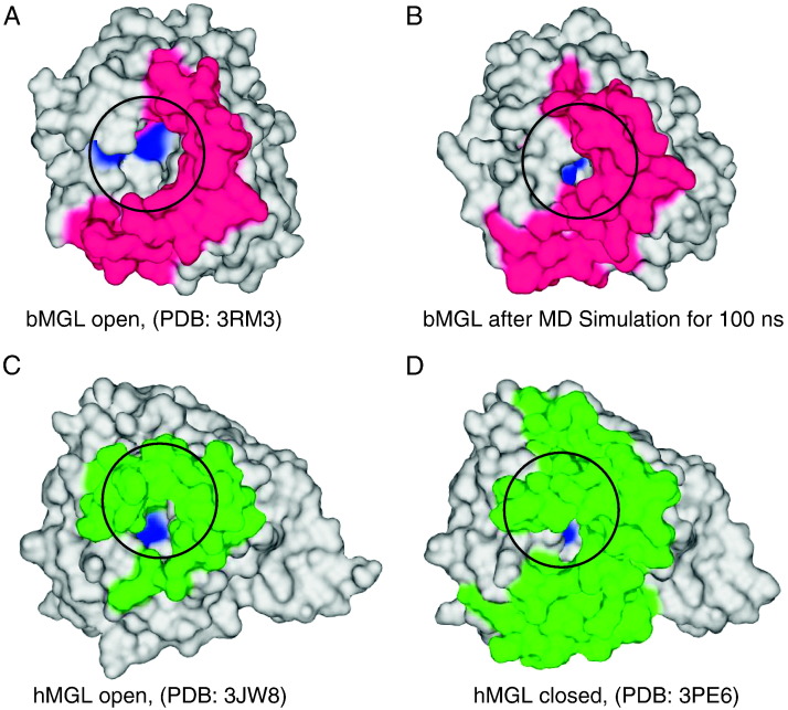
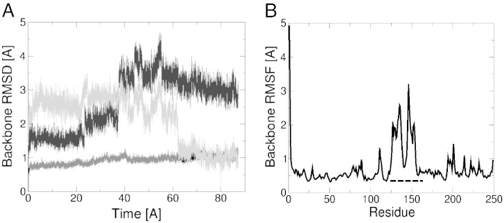
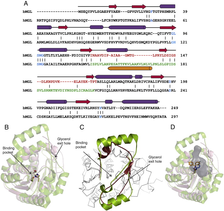
Monoacylglycerol lipases (MGLs) catalyse the hydrolysis of monoacylglycerol into free fatty acid and glycerol. MGLs have been identified throughout all genera of life and have adopted different substrate specificities depending on their physiological role. In humans, MGL plays an integral part in lipid metabolism affecting energy homeostasis, signalling processes and cancer cell progression. In bacteria, MGLs degrade short-chain monoacylglycerols which are otherwise toxic to the organism. We report the crystal structures of MGL from the bacterium Bacillus sp. H257 (bMGL) in its free form at 1.2Å and in complex with phenylmethylsulfonyl fluoride at 1.8Å resolution. In both structures, bMGL adopts an α/β hydrolase fold with a cap in an open conformation. Access to the active site residues, which were unambiguously identified from the protein structure, is facilitated by two different channels. The larger channel constitutes the highly hydrophobic substrate binding pocket with enough room to accommodate monoacylglycerol. The other channel is rather small and resembles the proposed glycerol exit hole in human MGL. Molecular dynamics simulation of bMGL yielded open and closed states of the entrance channel and the glycerol exit hole. Despite differences in the number of residues, secondary structure elements, and low sequence identity in the cap region, this first structure of a bacterial MGL reveals striking structural conservation of the overall cap architecture in comparison with human MGL. Thus it provides insight into the structural conservation of the cap amongst MGLs throughout evolution and provides a framework for rationalising substrate specificities in each organism.


Figures

Fig. 1
Fig. 1
Overall structure of monoacylglycerol lipase (MGL) from Bacillus sp. H257. The α/β hydrolase core is coloured wheat brown, the cap region (Ile119–Thr164) is represented in ruby red. A) The catalytic residues Ser97, Asp196, and His226 are represented as ball and sticks. The small α-helix in the cap region is labelled as α-cap; the letters N and C represent the respective termini of the protein. B) Tube representation of bMGL in the same orientation with the flexibility of individual residues indicated based on B-factor values.
Fig. 2
Fig. 2
Small angle X-ray scattering (SAXS) measurement of bMGL in solution. A) The theoretical scattering curve of the atomic model (black) shows a good fit to the experimental data (grey). B) Pair distribution functions estimated from the SAXS data (grey) and calculated for the crystal structure (black).
Fig. 3
Fig. 3
Surface and access paths to the catalytic site of bMGL. A and B) Surface representations of the uncomplexed bMGL structure showing the substrate-binding pocket and the glycerol exit hole in two different orientations. The colouring scheme represents the lipophilic potential: Regions coloured red are hydrophobic, blue are hydrophilic. A) Orientation of the protein as in Fig. 1. B) Orientation of the protein after rotation of 55 and 87° along X and Y axes, respectively, for better visualisation of the substrate binding pocket. C) Access paths to the active site of bMGL. The main active site channel is shown in blue, and the proposed glycerol exit hole is shown in green. Residues shown as red sticks are part of the catalytic triad (orientation of the molecule similar to Fig. 3B). D) Radii profile of the access paths of bMGL starting from the active site (Ser97) towards the protein surface.
Fig. 4
Fig. 4
Active site architecture of bMGL. A) The 2Fo–Fc sigma weighted electron density map (grey) is contoured at 1σ around the catalytic triad residues Ser97, Asp196 and His226 (violet sticks). B) The catalytic triad environment in the presence of a water molecule in the uncomplexed structure. H-bonds are depicted as dashed lines. The catalytic triad and oxyanion hole residues are shown in violet. The water molecule (W5) mediating the polar contacts is depicted in cyan. C) Environment of the catalytic triad in the bMGL–PMSF complex structure. H-bonds are depicted as dashed lines. The catalytic triad residues and oxyanion hole forming residues are shown in green. The phenyl group of the PMSF molecule covalently bound to Ser97 is represented in magenta. D) Depiction of atomic details of the interaction between bMGL and PMSF using LigPlot + .
Fig. 5
Fig. 5
Surface representation of open and closed conformations of human and bacterial MGL. A) Crystal structure of bMGL in the open conformation. B) bMGL structure in the closed conformation after a 100 ns unrestrained molecular dynamics simulation in explicit water. C) Crystal structure of hMGL in the open conformation and D) hMGL structure in the closed conformation with inhibitor bound. Active site residues are shown in blue, cap regions are shown in magenta and green for bMGL and hMGL respectively. The circles mark the entrance to the binding pocket.
Fig. 6
Fig. 6
Flexibility of individual residues in bMGL. A) Backbone root mean square deviation of bMGL H257 during a 90 ns molecular dynamics simulation. The backbone RMSD of the cap region relative to the open conformation is shown in black and relative to the closed confirmation in light grey, the core region of the protein is shown in dark grey (bottom line). B) Backbone root means square fluctuations (RMSF) per residue of bMGL during a 90 ns molecular dynamics simulation. The cap region from Ile119 to Thr164 is indicated with a dotted line.
Fig. 7
Fig. 7
Structural comparison of bacterial and human MGL. A) Structure based sequence alignment of bMGL and hMGL (PDB ID: 3HJU) using Dali . Secondary structure elements correspond to those observed in bMGL. β-strands and α-helices are depicted as red arrows and purple cylinders, respectively. Residues in blue correspond to the consensus G-X-S-X-G motif, the catalytic Asp and His, and the oxyanion hole forming residues. Residues within the cap regions of bMGL and hMGL are coloured in red and green, respectively. The proposed membrane binding helix α4 in the cap region of hMGL is shown as orange cylinder. B) Conservation of the catalytic triad evidenced by a structural superimposition. The structure of bMGL is shown as wheat brown cartoon and that of hMGL (PDB ID: 3JW8) as green cartoon. bMGL catalytic residues (Ser97-Asp196-His226) and hMGL (PDB ID: 3JW8) catalytic residues (Ser132-Asp249-His279) are shown as sticks in atomic colours. The substrate binding pocket and the proposed glycerol exit hole are indicated by arrows. C) Structural superposition of the cap region from bMGL (ruby red) and hMGL (green; PDB ID: 3JW8) shows a conserved architecture. D) bMGL (wheat brown) complexed to PMSF (blue) superimposed onto hMGL (green; PDB ID: 3JWE) complexed to SAR629 (orange). The cavity of bMGL as calculated by Casox is represented as a grey surface.

References

    1. Tornqvist H., Belfrage P. Purification and some properties of a monoacylglycerol-hydrolyzing enzyme of rat adipose tissue. J. Biol. Chem. 1976;251:813–819. - PubMed
    1. Fredrikson G., Tornqvist H., Belfrage P. Hormone-sensitive lipase and monoacylglycerol lipase are both required for complete degradation of adipocyte triacylglycerol. Biochim. Biophys. Acta. 1986;876:288–293. - PubMed
    1. Dinh T.P., Kathuria S., Piomelli D. RNA interference suggests a primary role for monoacylglycerol lipase in the degradation of the endocannabinoid 2-arachidonoylglycerol. Mol. Pharmacol. 2004;66:1260–1264. - PubMed
    1. Di Marzo V., Goparaju S.K., Wang L., Liu J., Batkai S., Jarai Z., Fezza F., Miura G.I., Palmiter R.D., Sugiura T., Kunos G. Leptin-regulated endocannabinoids are involved in maintaining food intake. Nature. 2001;410:822–825. - PubMed
    1. Hohmann A.G., Suplita R.L., Bolton N.M., Neely M.H., Fegley D., Mangieri R., Krey J.F., Walker J.M., Holmes P.V., Crystal J.D., Duranti A., Tontini A., Mor M., Tarzia G., Piomelli D. An endocannabinoid mechanism for stress-induced analgesia. Nature. 2005;435:1108–1112. - PubMed

Publication types

MeSH terms