. 2012 Jul;303(1):H75-85.
doi: 10.1152/ajpheart.00241.2012. Epub 2012 May 4.

Cardiac mTOR protects the heart against ischemia-reperfusion injury

Affiliations

Cardiac mTOR protects the heart against ischemia-reperfusion injury

Toshinori Aoyagi et al. Am J Physiol Heart Circ Physiol. 2012 Jul.

Abstract

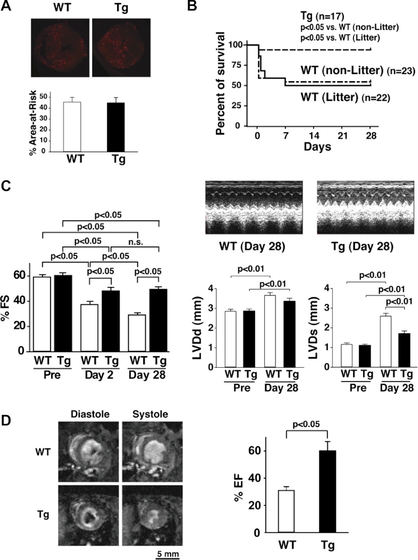
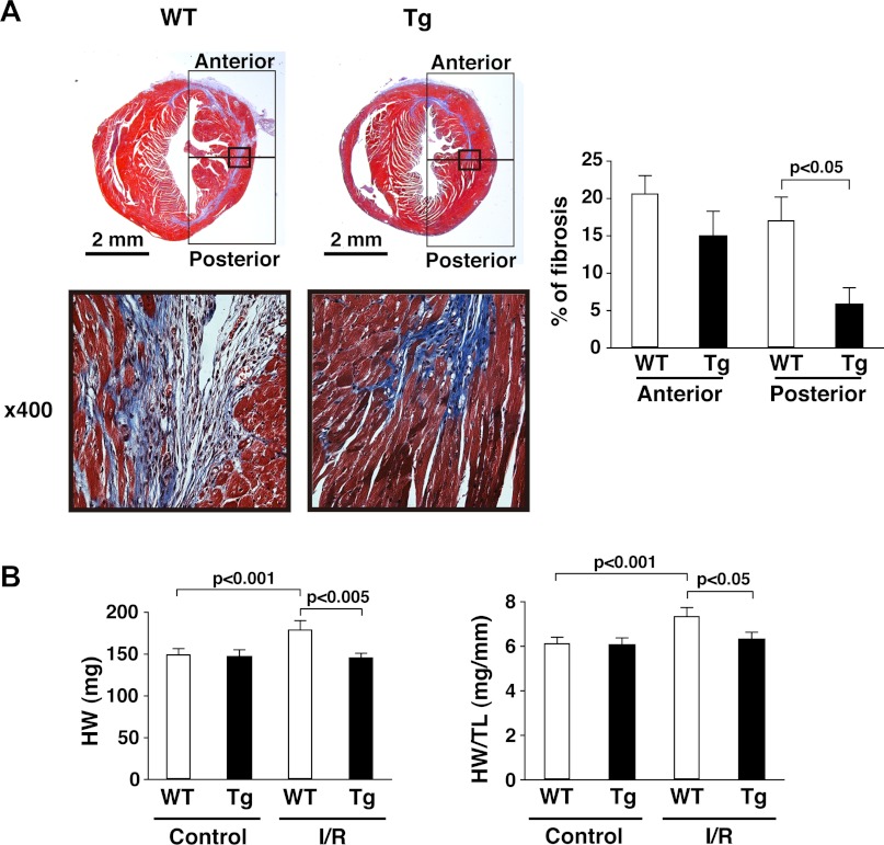
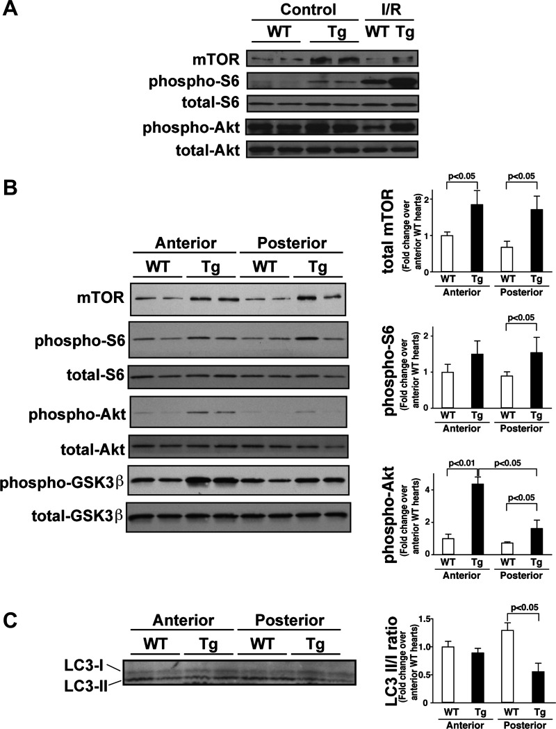
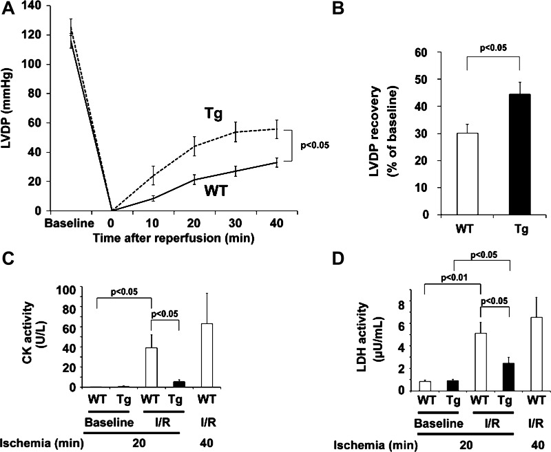
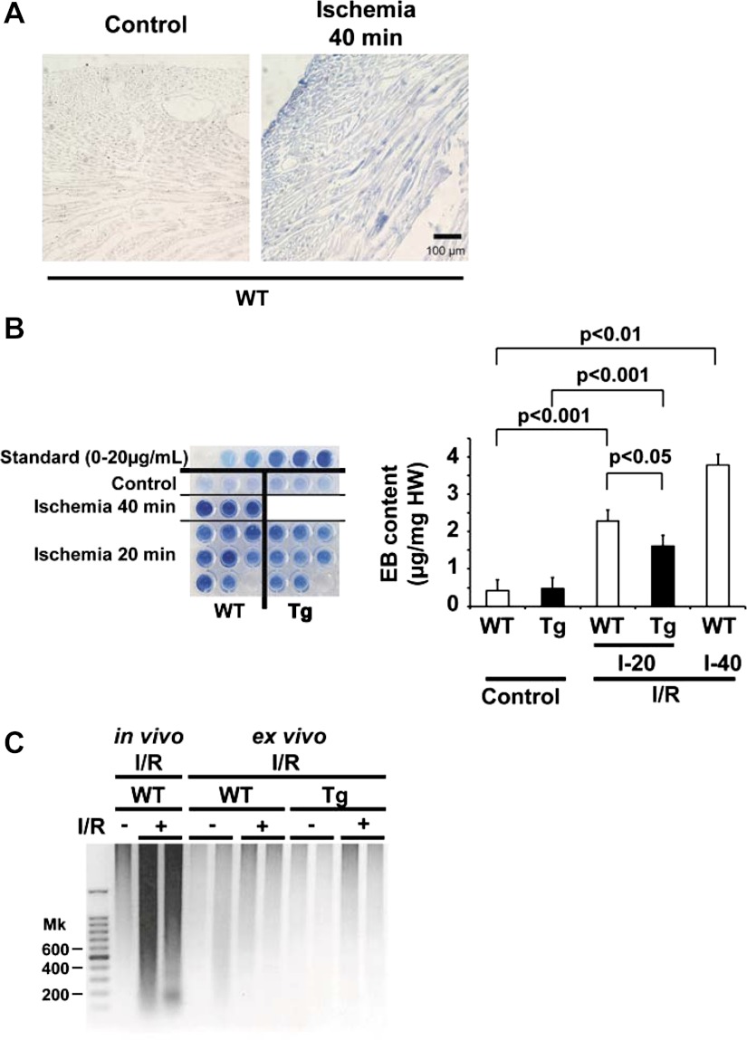
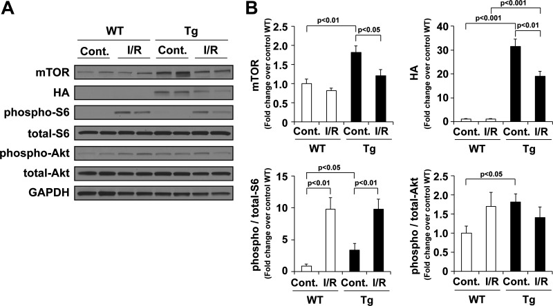
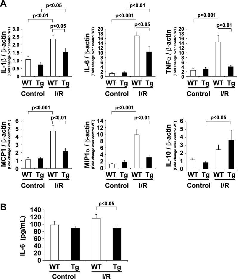
Cardiac mammalian target of rapamycin (mTOR) is necessary and sufficient to prevent cardiac dysfunction in pathological hypertrophy. However, the role of cardiac mTOR in heart failure after ischemic injury remains undefined. To address this question, we used transgenic (Tg) mice with cardiac-specific overexpression of mTOR (mTOR-Tg mice) to study ischemia-reperfusion (I/R) injury in two animal models: 1) in vivo I/R injury with transient coronary artery ligation and 2) ex vivo I/R injury in Langendorff-perfused hearts with transient global ischemia. At 28 days after I/R, mortality was lower in mTOR-Tg mice than littermate control mice [wild-type (WT) mice]. Echocardiography and MRI demonstrated that global cardiac function in mTOR-Tg mice was preserved, whereas WT mice exhibited significant cardiac dysfunction. Masson's trichrome staining showed that 28 days after I/R, the area of interstitial fibrosis was smaller in mTOR-Tg mice compared with WT mice, suggesting that adverse left ventricular remodeling is inhibited in mTOR-Tg mice. In the ex vivo I/R model, mTOR-Tg hearts demonstrated improved functional recovery compared with WT hearts. Perfusion with Evans blue after ex vivo I/R yielded less staining in mTOR-Tg hearts than WT hearts, indicating that mTOR overexpression inhibited necrosis during I/R injury. Expression of proinflammatory cytokines, including IL-6 and TNF-α, in mTOR-Tg hearts was lower than in WT hearts. Consistent with this, IL-6 in the effluent post-I/R injury was lower in mTOR-Tg hearts than in WT hearts. These findings suggest that cardiac mTOR overexpression in the heart is sufficient to provide substantial cardioprotection against I/R injury and suppress the inflammatory response.


Figures

Fig. 1.
Fig. 1.
Mammalian target of rapamycin (mTOR) overexpression protects the heart and preserves cardiac function after in vivo ischemia-reperfusion (I/R). A: the ischemic area (area at risk). Top, representative images of fluorescent microspheres from wild-type (WT) and mTOR-transgenic (Tg) mice. Hearts were harvested at 28 days after I/R. Bottom, percentage of the area at risk of hearts from WT and mTOR-Tg mice. The area at risk was assessed as previously described (22). n = 5 mice/group. B: Kaplan-Meier survival curves from nonlittermate (non-Litter) WT, littermate (Litter) WT, and mTOR-Tg mice after I/R with temporal coronary ligation. n = 17 mTOR-Tg mice, 23 nonlittermate WT mice, and 22 littermate WT mice. C: echocardiographic analyses. Right, representative M-mode images of operated WT and mTOR-Tg mice at 28 days after I/R as well as left ventricular (LV) diastolic dimension (LVDd) and LV systolic dimension (LVDs) at pre-I/R and 28 days after I/R. Left, mean scores for percent fractional shortening (%FS) at pre-I/R and 2 and 28 days after I/R. n = 11 WT mice and 16 mTOR-Tg mice. n.s., Not significant. D: MRI of the heart at 28 days after I/R. Left, representative pair of images from littermate pairs of male mTOR-Tg and WT mice. Right, percent ejection fraction (%EF) calculated from MRI images. n = 3 WT mice and 5 mTOR-Tg mice.
Fig. 2.
Fig. 2.
mTOR overexpression suppresses adverse LV remodeling. A: interstitial myocardial fibrosis. Left, representative photos of Masson's trichrome staining in cardiac sections from WT and mTOR-Tg mice at 28 days after I/R. Bottom, magnified images of the regions indicated by the squares in each heart section of the top images. Right, quantitative analysis of interstitial fibrosis examined by Masson's trichrome staining. n = 5 mice/group. B: heart weight (HW; left) and ratios of HW to tibia length (TL; right). Hearts from each group were harvested after echocardiography was performed. n = 11 control WT mice, 10 control mTOR-Tg mice, 11 I/R WT mice, and 16 I/R mTOR-Tg mice.
Fig. 3.
Fig. 3.
mTOR-related signaling pathways and autophagy in mTOR-Tg mice after in vivo I/R. A: representative immunoblots of mTOR signaling molecules in the whole heart. Hearts were harvested at 28 days after I/R. Control hearts were harvested from sham-operated mice. Immunoblot analysis was performed with the indicated antibodies. Blots are representative of three independent samples in each group. B: immunoblots of mTOR signaling molecules in cardiac tissues from either the anterior or posterior LV wall. Left, representative immunoblots of mTOR signaling molecules in the anterior and posterior LV wall. The heart was harvested at 28 days after I/R, and the LV wall was split into anterior and posterior sections to produce lysates for Western blot analysis. Right, densitometric quantification of immunoblots. GSK, glycogen synthase kinase. C: analysis of autophagy. Left, representative immunoblots of LC3-I and LC3-II in the anterior and posterior LV wall. Right, ratio of LC3-II to LC3-I (LC3-II/I). Data were normalized to the mean protein level in the anterior LV of WT hearts. n = 5 WT mice and 3 mTOR-Tg mice.
Fig. 4.
Fig. 4.
mTOR overexpression preserves cardiac function and prevents cardiac injury during ex vivo I/R. A: LV developed pressure (LVDP) profiles during I/R in WT and mTOR-Tg mice. n = 28 WT mice and 19 mTOR-Tg mice. B: maximum LVDP recovery (percentage of baseline) measured at 40 min of reperfusion. n = 28 WT mice and 19 mTOR-Tg mice. C and D: activity of creatine kinase (CK) and lactate dehydrogenase (LDH) in the effluent collected during the reperfusion period. To determine enzyme activities immediately after ex vivo I/R injury, effluents from hearts exposed to either 20 or 40 min of global ischemia were collected at control perfusion (baseline) and after 40-min reperfusion (I/R). n = 7 WT mice and 8 mTOR-Tg mice in hearts subjected to 20-min ischemia, and n = 3 WT mice in hearts subjected to 40-min ischemia.
Fig. 5.
Fig. 5.
mTOR overexpression prevents necrosis in ex vivo I/R injury. A: representative tissue sections from hearts perfused with Evans blue (EB). Hearts subjected to 15-min control perfusion or 40-min ischemia/40-min reperfusion (Ischemia 40 min) in WT mice were harvested after perfusion with EB. B, left: EB dye extracted from cardiac tissue. Standard samples (0–20 μg/ml) are shown at the top. WT hearts from 15-min control perfusion (n = 3), mTOR-Tg hearts from 15-min control perfusion (n = 3), WT hearts from 40-min ischemia/40-min reperfusion (I-40; n = 3), and WT (n = 8) and mTOR-Tg (n = 8) hearts from 20-min ischemia/40-min reperfusion (I-20) were examined for this assay. Right, quantification of EB incorporation in the myocardium. The color intensity of the samples on the left was measured as described in methods. C: DNA laddering. Genomic DNA was prepared from hearts subjected to either in vivo or ex vivo I/R injury. Twenty-eight days post-I/R, cardiac tissue from hearts subjected to in vivo I/R injury was harvested at the border zone of the LV, where significant apoptosis has been observed after I/R injury (40). Cardiac tissue from hearts subjected to ex vivo I/R injury was harvested from the LV wall after 20-min ischemia/40-min reperfusion. The DNA laddering shown is representative of results from 3 independent samples/group.
Fig. 6.
Fig. 6.
mTOR signaling pathways in mTOR-Tg mice after ex vivo I/R. A: representative immunoblots of mTOR signaling molecules in hearts subjected to ex vivo Langendorff perfusion. Hearts were harvested after 20-min ischemia/40-min reperfusion ex vivo. Control hearts were harvested at 15-min control perfusion. Immunoblot analysis was performed with the indicated antibodies. Blots are representative of 4 independent samples/group. B: quantitative analysis of mTOR, hemagglutinin (HA), phospho-S6, and phospho-Akt. Values were normalized to levels in control WT hearts in each experiment. n = 8 mice/group.
Fig. 7.
Fig. 7.
Inflammatory response in mTOR-Tg mice after ex vivo I/R. A: expression of IL-1β, IL-6, TNF-α, monocyte chemotactic protein (MCP)-1, macrophage inflammatory protein (MIP)-1α, and IL-10 mRNA levels in the heart. mRNA was measured by quantitative RT-PCR. n = 6–9 mice/group. B: IL-6 protein secreted from ex vivo perfused hearts. Effluents were collected at 15-min control perfusion and after 20-min ischemia/40-min reperfusion (I/R) in each group. n = 27 WT mice and 19 mTOR-Tg mice.

References

    1. Avruch J, Hara K, Lin Y, Liu M, Long X, Ortiz-Vega S, Yonezawa K. Insulin and amino-acid regulation of mTOR signaling and kinase activity through the Rheb GTPase. Oncogene 25: 6361–6372, 2006 - PubMed
    1. Baines CP, Kaiser RA, Purcell NH, Blair NS, Osinska H, Hambleton MA, Brunskill EW, Sayen MR, Gottlieb RA, Dorn GW, Robbins J, Molkentin JD. Loss of cyclophilin D reveals a critical role for mitochondrial permeability transition in cell death. Nature 434: 658–662, 2005 - PubMed
    1. Burchfield JS, Dong JW, Sakata Y, Gao F, Tzeng HP, Topkara VK, Entman ML, Sivasubramanian N, Mann DL. The cytoprotective effects of tumor necrosis factor are conveyed through tumor necrosis factor receptor-associated factor 2 in the heart. Circ Heart Fail 3: 157–164, 2010 - PMC - PubMed
    1. Buss SJ, Muenz S, Riffel JH, Malekar P, Hagenmueller M, Weiss CS, Bea F, Bekeredjian R, Schinke-Braun M, Izumo S, Katus HA, Hardt SE. Beneficial effects of Mammalian target of rapamycin inhibition on left ventricular remodeling after myocardial infarction. J Am Coll Cardiol 54: 2435–2446, 2009 - PubMed
    1. Cittadini A, Monti MG, Petrillo V, Esposito G, Imparato G, Luciani A, Urciuolo F, Bobbio E, Natale CF, Sacca L, Netti PA. Complementary therapeutic effects of dual delivery of insulin-like growth factor-1 and vascular endothelial growth factor by gelatin microspheres in experimental heart failure. Eur J Heart Fail 13: 1264–1274, 2011 - PubMed

Publication types

MeSH terms

LinkOut - more resources