

Subharmonic microbubble emissions for noninvasively tracking right ventricular pressures
- PMID: 22561300
- PMCID: PMC3404645
- DOI: 10.1152/ajpheart.00560.2011
Subharmonic microbubble emissions for noninvasively tracking right ventricular pressures
Abstract
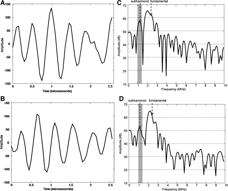
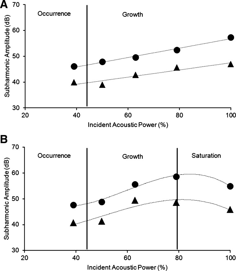
Right heart catheterization is often required to monitor intra-cardiac pressures in a number of disease states. Ultrasound contrast agents can produce pressure modulated subharmonic emissions that may be used to estimate right ventricular (RV) pressures. A technique based on subharmonic acoustic emissions from ultrasound contrast agents to track RV pressures noninvasively has been developed and its clinical potential evaluated. The subharmonic signals were obtained from the aorta, RV, and right atrium (RA) of five anesthetized closed-chest mongrel dogs using a SonixRP ultrasound scanner and PA4-2 phased array. Simultaneous pressure measurements were obtained using a 5-French solid state micromanometer tipped catheter. Initially, aortic subharmonic signals and systemic blood pressures were used to obtain a calibration factor in units of millimeters of mercury per decibel. This factor was combined with RA pressures (that can be obtained noninvasively) and the acoustic data from the RV to obtain RV pressure values. The individual calibration factors ranged from -2.0 to -4.0 mmHg/dB. The subharmonic signals tracked transient changes in the RV pressures within an error of 0.6 mmHg. Relative to the catheter pressures, the mean errors in estimating RV peak systolic and minimum diastolic pressures, and RV relaxation [isovolumic negative derivative of change in pressure over time (-dP/dt)] by use of the subharmonic signals, were -2.3 mmHg, -0.8 mmHg, and 2.9 mmHg/s, respectively. Overall, acoustic estimates of RV peak systolic and minimum diastolic pressures and RV relaxation were within 3.4 mmHg, 1.8 mmHg, and 5.9 mmHg/s, respectively, of the measured pressures. This pilot study demonstrates that subharmonic emissions from ultrasound contrast agents have the potential to noninvasively track in vivo RV pressures with errors below 3.5 mmHg.
Figures

References
-
- Andersen K, Jensen J. Ambient pressure sensitivity of microbubbles investigated through a parameter study. J Acoust Soc Am 126: 3350–3358, 2009 - PubMed
-
- Asch FM, Weissman NJ. Overview of the 2008 Food and Drug Administration advisory committee on safety consideration in the development of ultrasound contrast agents. Circulation 119: 1956–1961, 2009 - PubMed
-
- Avi-Mor V, Sugeng L, Lindner JR. Imaging the forgotten chamber: is the devil in the boundary? J Am Soc Echocardiogr 23: 141–143, 2010 - PubMed
-
- Bouakaz A, Frinking P, de Jong N, Bom N. Noninvasive measurement of the hydrostatic pressure in a fluid filled cavity based on the disappearance time of micrometer-sized free gas bubbles. Ultrasound Med Biol 25: 1407–1415, 1999 - PubMed
Publication types
MeSH terms
Substances
Grants and funding
LinkOut - more resources
Full Text Sources
Other Literature Sources
Medical
Miscellaneous