

Epithelial-mesenchymal transition, TGF-β, and osteopontin in wound healing and tissue remodeling after injury
- PMID: 22561306
- PMCID: PMC5207477
- DOI: 10.1097/BCR.0b013e318240541e
Epithelial-mesenchymal transition, TGF-β, and osteopontin in wound healing and tissue remodeling after injury
Abstract
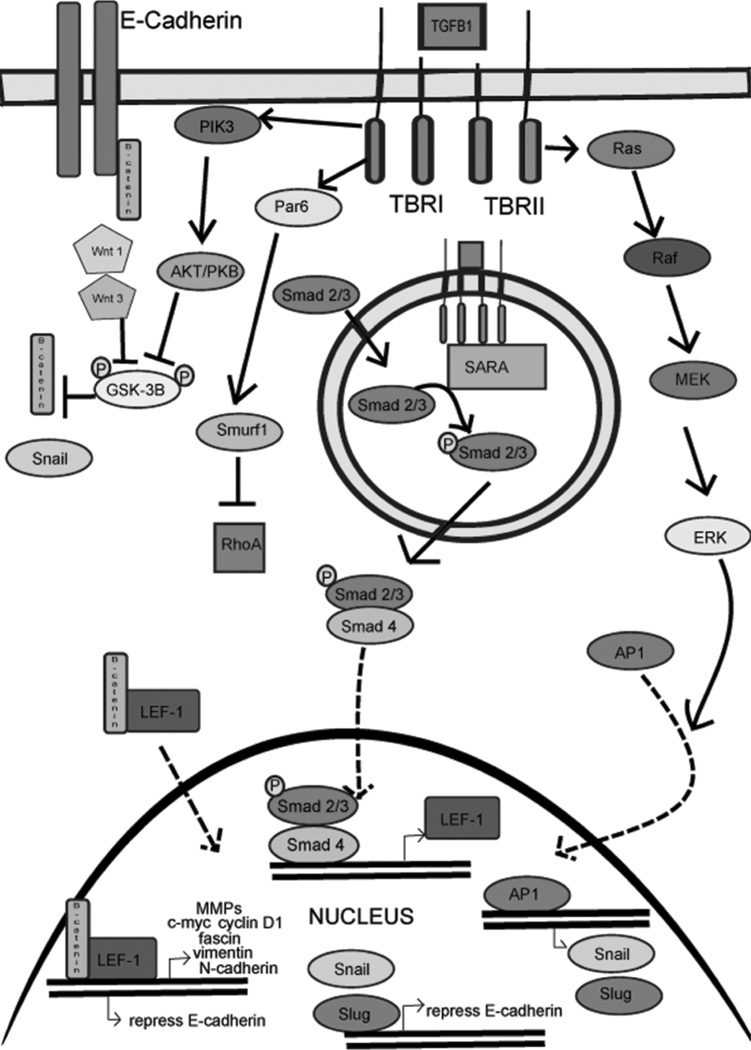
Epithelial-mesenchymal transition (EMT) is a process essential to wound healing and tissue remodeling after a thermal burn or other injury. EMT is characterized by phenotypic changes in epithelial cells that render them apolar, with decreased cell-cell adhesions, increased motility, and changes in cytoskeletal architecture similar to mesenchymal stem cells. With regard to healing a thermal burn wound, many facets of wound healing necessitate cells to undergo these phenotypic changes; two will be described in the following review. The first is the differentiation of epithelial cells into myofibroblasts that rebuild the extracellular matrix and facilitate wound contraction. The second is reepithelialization by keratinocytes. The primary cytokine signal identified in the literature that triggers EMT is transforming growth factor (TGF)-β. In addition to its vital role in the induction of EMT, TGF-β has many other roles in the wound healing process. The following review will provide evidence that EMT is a central event in wound healing. It will also show the importance of a regulated amount of TGF-β for proper wound healing. Finally, osteopontin will be briefly discussed with its relation to wound healing and its connections to EMT and TGF-β.
Figures

References
Publication types
MeSH terms
Substances
Grants and funding
LinkOut - more resources
Full Text Sources
Other Literature Sources
Medical
Research Materials
