. 2012 Sep 21;30(43):6216-24.
doi: 10.1016/j.vaccine.2012.04.077. Epub 2012 May 1.

Lymphocyte-polarized dendritic cells are highly effective in inducing tumor-specific CTLs

Affiliations

Lymphocyte-polarized dendritic cells are highly effective in inducing tumor-specific CTLs

Erik Berk et al. Vaccine. .

Abstract

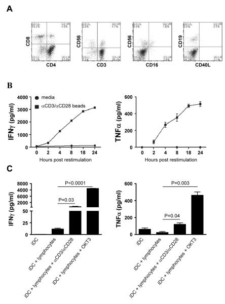
High activity of dendritic cells (DCs) in inducing cytotoxic T cells (CTLs) led to their application as therapeutic cancer vaccines. The ability of DCs to produce IL-12p70 is one of the key requirements for effective CTL induction and a predictive marker of their therapeutic efficacy in vivo. We have previously reported that defined cocktails of cytokines, involving TNFα and IFNγ, induce mature type-1 polarized DCs (DC1s) which produce strongly elevated levels of IL-12 and CXCL10/IP10 upon CD40 ligation compared to "standard" PGE₂-matured DCs (sDCs; matured with IL-1β, IL-6, TNFα, and PGE₂) and show higher CTL-inducing activity. Guided by our observations that DC1s can be induced by TNFα- and IFNγ-producing CD8⁺ T cells, we have tested the feasibility of using lymphocytes to generate DC1s in a clinically-compatible process, to limit the need for clinical-grade recombinant cytokines and the associated costs. CD3/CD28 activation of bulk lymphocytes expanded them and primed them for effective production of IFNγ and TNFα following restimulation. Restimulated lymphocytes, or their culture supernatants, enhanced the maturation status of immature (i)DCs, elevating their expression of CD80, CD83 and CCR7, and the ability to produce IL-12p70 and CXCL10 upon subsequent CD40 ligation. The "lymphocyte-matured" DC1s showed elevated migration in response to the lymph-node-directing chemokine, CCL21, when compared to iDCs. When loaded with antigenic peptides, supernatant-matured DCs induced much high levels of CTLs recognizing tumor-associated antigenic epitope, than PGE₂-matured DCs from the same donors. These results demonstrate the feasibility of generation of polarized DC1s using autologous lymphocytes.


Figures

Figure 1
Figure 1. Ex vivo-expanded lymphocytes rapidly produce IFNγand TNFαupon restimulation
Isolated lymphocytes were expanded for 7 days with αCD3- and αCD28-coated beads. A) The phenotype of day 7 expanded lymphocytes was determined by flow cytometry. B) Day 7 expanded lymphocytes were harvested and either cultured in the absence of additional stimulation (circles) or restimulated (squares). At the indicated times, supernatants were collected to determine the secretion of IFNγ(left) and TNFα (right). C) Autologous day 5 iDCs were cultured in the presence of the day 7 expanded lymphocytes in the presence or absence of the indicated stimuli. After 24 hours of co-cultures, supernatants were collect and the production of IFNγ(left) and TNFα(right) was determined. Data shown from one of at least 2 independent experiments that yielded similar results.
Figure 2
Figure 2. Restimulated expanded lymphocytes or supernatant induce the maturation of autologous DCs and primes them for high IL-12p70 and CXCL10 production
iDC were cultured for 24 hours with autologous day 7 expanded lymphocytes in the presence or absence of the indicated stimuli or with supernatant from 24 hours restimulated lymphocytes. A) After 24 hours, the morphology of the DCs was analyzed using bright field microscopy. The morphology of lymphocyte-matured DCs was compared with that of iDCs and with that of DCs exposed to the non-polarizing maturation-inducing cytokine cocktail (PGE2-matured sDC: IL1β, TNFα, IL-6 and PGE2). B) 24 hour matured DCs were collected, washed and rested overnight at 37°C in fresh media to remove any CCL19 produced during maturation and allow for re-expression of chemokine receptor CCR7 on the cell surface [26] (see M&M). After resting, the phenotype of the DCs was determined by flow cytometry. C) Average Mean Fluorescence Intensity expression of DC maturation markers of 3-5 individual experiments. D-E) 24 hour matured DCs were collected, washed and subsequently cultured for 24 hour with CD40L-expressing J558 cells (2×10⁴ DC: 5×10⁴ J558). After co-culture, supernatant was collected and analyzed for the presence of (D) IL12p70 and (E) CXCL10. Data shown are representative results from at least 2 independent experiments. F) Comparison of average of Mean Fluorescence Intensity of DC maturation associated markers on lymphocyte-matured DCs from 4 healthy donors with those of 3 melanoma patients. G) Comparison of IL-12p70 production by immature and lymphocyte-matured DCs from healthy donors (4) and melanoma patients (3) after 24 hours of CD40L stimulation.
Figure 3
Figure 3. Supernatant-matured DCs efficiently migrate in response to CCL21
24 hour matured DCs were collected, washed and rested overnight at 37°C. After resting, DCs were placed in the top chamber of a transwell system and allowed to migrate for 3 hours towards CCL21 (in the bottom chamber) in a chemotaxis assay. Following incubation, the migrated cells in the bottom chamber of the transwell were collected and counted using a flow cytometer (see M&M). Migrated cells are represented as percentage of total cells. Migrated cells were counted twice and data is shown as mean +/− SD. Data shown are representative results from 2 independent experiments.
Figure 4
Figure 4. Tumor-peptide-loaded supernatant-matured DCs induce strong anti-tumor CTL responses from autologous naïve CD8+ T cells
24 hour-matured DCs (supernatant-matured or PGE2-matured sDC) from healthy donors were collected, washed and loaded for 2 hours with tumor peptides (MART-1, gp100, PAP3, and PSA2). Following peptide loading, DCs were cultured with autologous naïve CD8+ T cells at a 1:10 ratio. A) On day 12, CD8+ T cells were collected and the percentage of MART-1-specifc CD8+ T cells was determined by tetramer staining. B) Day 12 CD8+ T cells were collected, washed and used in a 24 hour IFNγ-ELISPOT analysis against tumor-peptides. Data are shown as mean +/− SEM of triplicate spot counts of one of 3 independent experiments that yielded similar results.

Similar articles

Cited by

References

    1. Lanzavecchia A, Sallusto F. Regulation of T cell immunity by dendritic cells. Cell. 2001 Aug 10;106(3):263–266. - PubMed
    1. Steinman RM. The dendritic cell system and its role in immunogenicity. Annu Rev Immunol. 1991;9:271–296. - PubMed
    1. Banchereau J, Briere F, Caux C, Davoust J, Lebecque S, Liu YJ, Pulendran B, Palucka K. Immunobiology of dendritic cells. Annu Rev Immunol. 2000;18:767–811. - PubMed
    1. Nestle FO, Filgueira L, Nickoloff BJ, Burg G. Human dermal dendritic cells process and present soluble protein antigens. J Invest Dermatol. 1998 May;110(5):762–766. - PubMed
    1. Kalinski P, Hilkens CM, Wierenga EA, Kapsenberg ML. T-cell priming by type-1 and type-2 polarized dendritic cells: the concept of a third signal. Immunol Today. 1999 Dec;20(12):561–567. - PubMed

Publication types

MeSH terms