. 2012 Jul:100:1-6.
doi: 10.1016/j.exer.2012.04.005. Epub 2012 Apr 24.

In vivo detection of experimental optic neuritis by pupillometry

Affiliations

In vivo detection of experimental optic neuritis by pupillometry

Kenneth S Shindler et al. Exp Eye Res. 2012 Jul.

Abstract

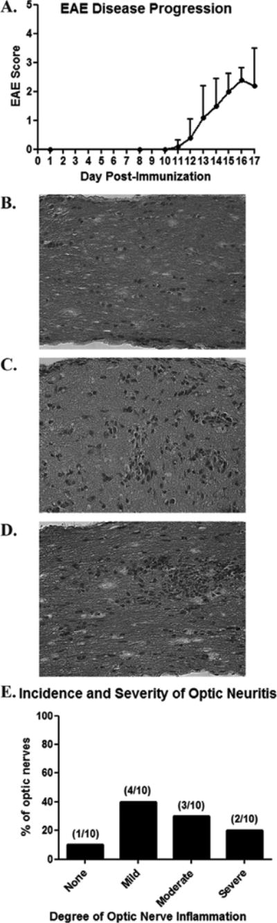
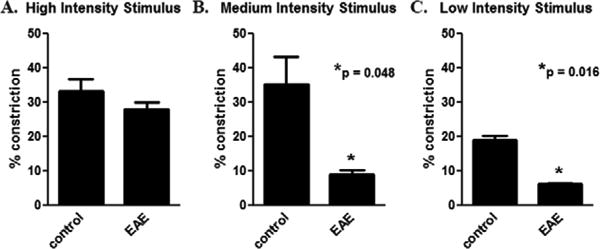
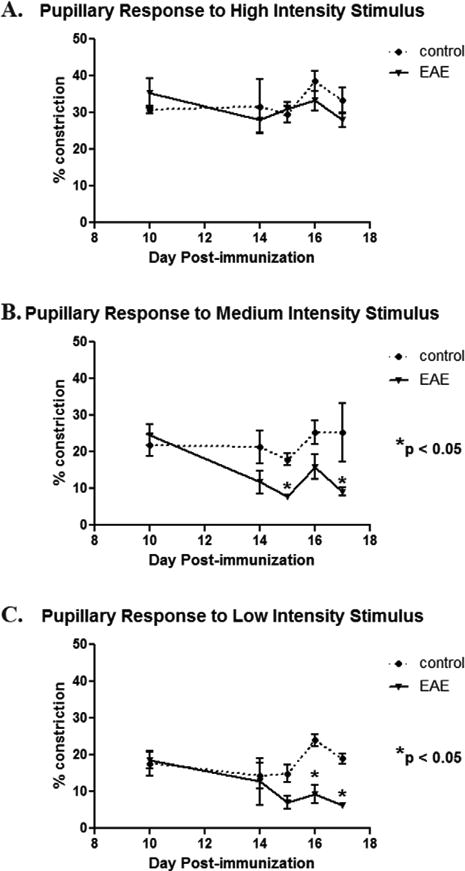
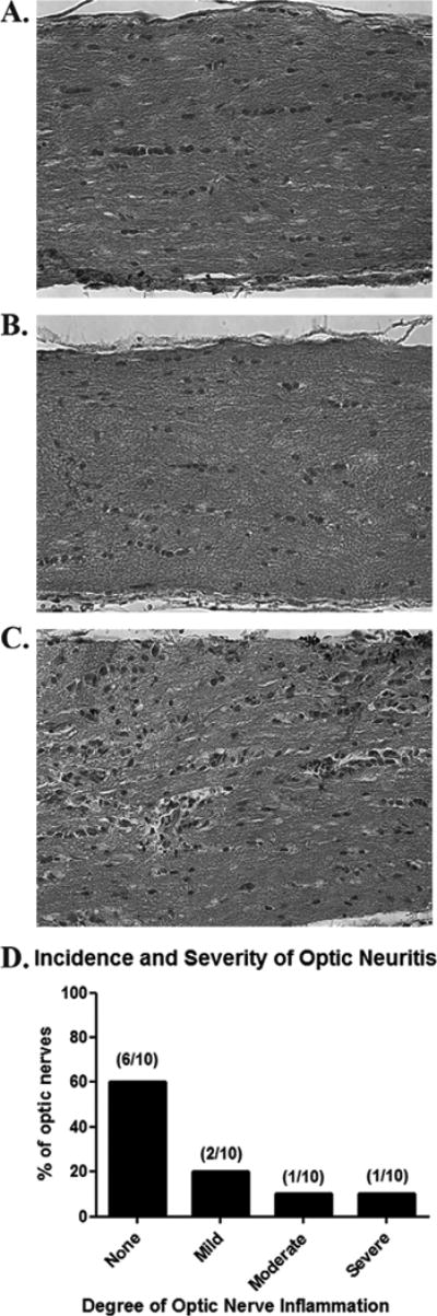
Optic neuritis is an inflammatory demyelination of optic nerve often occurring in multiple sclerosis (MS) patients. Mice with experimental autoimmune encephalomyelitis (EAE), an MS model, develop optic neuritis, but it is detected histologically after sacrifice, limiting the ability to monitor progression or treatment in vivo. We examined whether pupillary light responses measured by pupillometry can identify eyes with optic neuritis in EAE mice. C57BL/6 mice were exposed to unilateral light flashes of increasing intensity at 10 s intervals (4.7, 37, and 300 μW/cm(2)). Pupillary responses were recorded with a commercially available pupillometer. EAE was then induced by immunization with myelin oligodendrocyte glycoprotein. Pupillometry was repeated up to 17 days post-immunization, and responses were correlated with optic nerve inflammation. By day 17 post-immunization, 90% of EAE eyes had optic nerve inflammation. EAE eyes had significantly reduced pupillary constriction compared to control eyes. Mice exhibited more than a 25% decrease in pupillary constriction in at least one eye by days 13-15 post-immunization. In some eyes, pupil responses decreased prior to onset of detectable inflammation. Results show that pupillometry detects decreased optic nerve function in experimental optic neuritis, even in the absence of histological detection. Measuring pupillary constriction allows in vivo identification and functional assessment of eyes with optic neuritis that will be useful in evaluating potential therapies over time. Furthermore, results demonstrate that decreased visual function occurs early in optic neuritis, before optic nerve inflammation reaches its peak level.


Figures

Fig. 1
Fig. 1
High incidence of optic neuritis occurs in chronic murine EAE. C57BL/6 mice were immunized on day 0 with MOG to induce EAE. (A) Clinical tail and limb paralysis, hallmarks of EAE inflammation in spinal cord, began by days 12-13 post-immunization in all mice (n = 5), with severity increasing through at least day 15. (B) Longitudinal section of an optic nerve from a control, non-EAE mouse stained by H&E shows the normal cellularity. (C) An optic nerve from a day 17 post-immunization EAE mouse demonstrates mild inflammation, with small scattered foci of increased cellularity. (D) Moderate inflammation, with a focal area containing marked cellular infiltrate, is shown. (E) Histological examination of 10 EAE optic nerves 17 days post-inflammation demonstrates 90% of nerves develop optic neuritis, with varying degrees of inflammation. Original magnification X40 (B-D).
Fig. 2
Fig. 2
Pupillary light responses are reduced in EAE eyes. Pupillometry was performed in C57BL/6 mice before immunization and again 17 days later, after onset of EAE and optic neuritis. Pupil responses are recorded as the average percentage area constriction (pupil area prior to light stimulus – small pupil size post-stimulus/pupil area prior to light stimulus) in response to a single light flash in each eye (n = 10). (A) A 300 μW/cm2 light stimulus induced a 33.2 ± 3.5% constriction of the pupils in control mice, with no significant difference in EAE mice. (B) A 37 μW/cm2 light stimulus induced a 35.3 ± 8.1% constriction of the pupils in control mice, and a significantly smaller 9.1 ± 1.2% constriction in EAE mice. (C) A 4.7 μW/cm2 light stimulus induced an 18.9 ± 1.5% constriction of the pupils in control mice, and a significantly smaller 6.2 ± 0.4% constriction in EAE mice.
Fig. 3
Fig. 3
Pupillary light responses decrease by day 15 post-immunization in EAE mice. Pupillometry was repeated on multiple days after immunization with MOG to induce EAE in C57BL/6 mice. (A) Similar to findings at day 17, no difference in the degree of pupillary constriction induced by a 300 μW/cm2 light stimulus was found at any time point between control and EAE mouse eyes (n = 10). (B) Pupil constriction in response to a 37 μW/cm2 stimulus was no different between control and EAE eyes at early time points, with decreasing pupillary responses developing gradually in EAE mice and reaching statistical significance at day 15 post-immunization. (C) Similar to the medium intensity stimulus, pupil constriction in response to a 4.7 μW/cm2 stimulus was significantly decreased by day 16 post-immunization in EAE eyes as compared to control eyes.
Fig. 4
Fig. 4
Decreased pupillary light responses precede histological evidence of optic neuritis. Baseline pupillometry was performed using a 4.7 μW/cm2 stimulus prior to induction of EAE, and was repeated daily beginning 10 days post-immunization. Mice (n = 5) were sacrificed on the first day that one or both eyes demonstrated a 25% or greater decrease in pupillary constriction compared to baseline. (A,B) Normal optic nerve histology, without evidence of inflammation, is shown from two representative EAE mouse eyes that had marked (greater than 25%) decreases in pupillary light response. (C) Mild inflammation observed in another EAE eye that had decreased pupillary constriction. (D) While 8 of 10 eyes from EAE mice had 25% or greater decrease in pupillary constriction at the time of sacrifice, only 4 eyes demonstrated varying levels of inflammation on histological evaluation, with 60% of eyes exhibiting no detectable inflammation. Original magnification X40 (A-C).

References

    1. Arnold AC. Evolving management of optic neuritis and multiple sclerosis. Am J Ophthalmol. 2005;139:1101–1108. - PubMed
    1. Beck RW, Cleary PA, Anderson MM, Jr, Keltner JL, Shults WT, Kaufman DI, Buckley EG, Corbett JJ, Kupersmith MJ, Miller NR, et al. A randomized, controlled trial of corticosteroids in the treatment of acute optic neuritis. New Eng J Med. 1992;326:581–588. - PubMed
    1. Dutt M, Tabuena P, Ventura E, Rostami AM, Shindler KS. Timing of corticosteroid therapy is critical to prevent retinal ganglion cell loss in experimental optic neuritis. Invest Ophthalmol Vis Sci. 2010;51:1439–1445. - PMC - PubMed
    1. Ellis CJ. The afferent pupillary defect in acute optic neuritis. J Neurol Neurosurg Psychiatry. 1979;42:1008–1017. - PMC - PubMed
    1. Guy J, Qi X, Hauswirth WW. Adeno-associated viral-mediated catalase expression suppresses optic neuritis in experimental allergic encephalomyelitis. Proc Natl Acad Sci. 1998;95:13847–13852. - PMC - PubMed

Publication types

Substances