. 2012 Aug;40(14):6966-77.
doi: 10.1093/nar/gks369. Epub 2012 May 4.

The structure of the C-terminal domain of the largest editosome interaction protein and its role in promoting RNA binding by RNA-editing ligase L2

Affiliations

The structure of the C-terminal domain of the largest editosome interaction protein and its role in promoting RNA binding by RNA-editing ligase L2

Young-Jun Park et al. Nucleic Acids Res. 2012 Aug.

Abstract

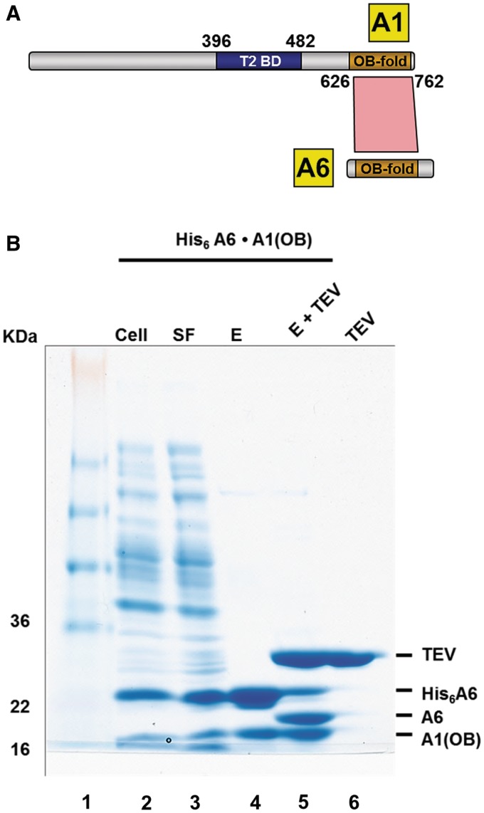
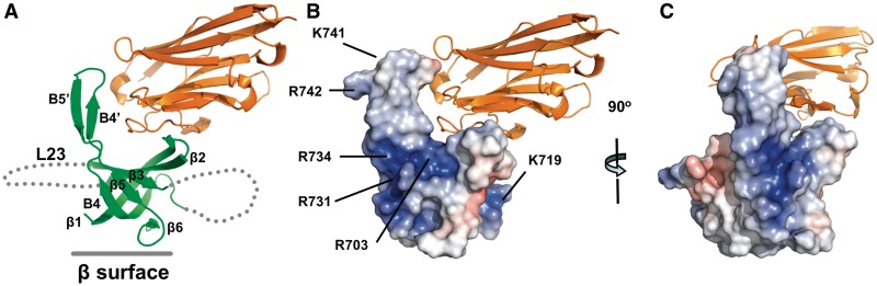
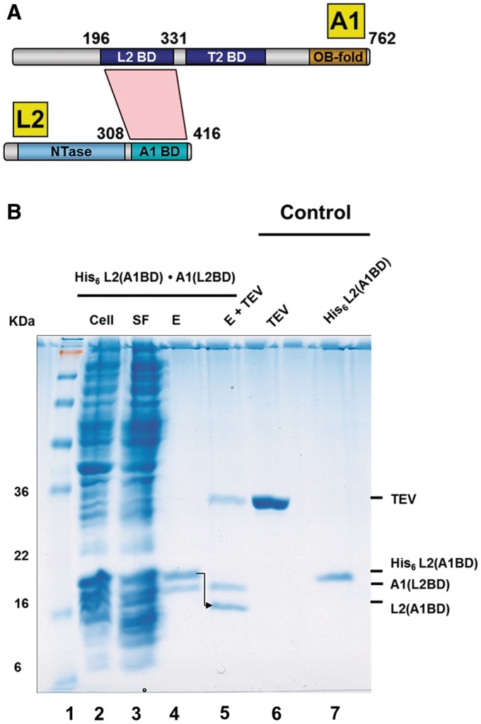
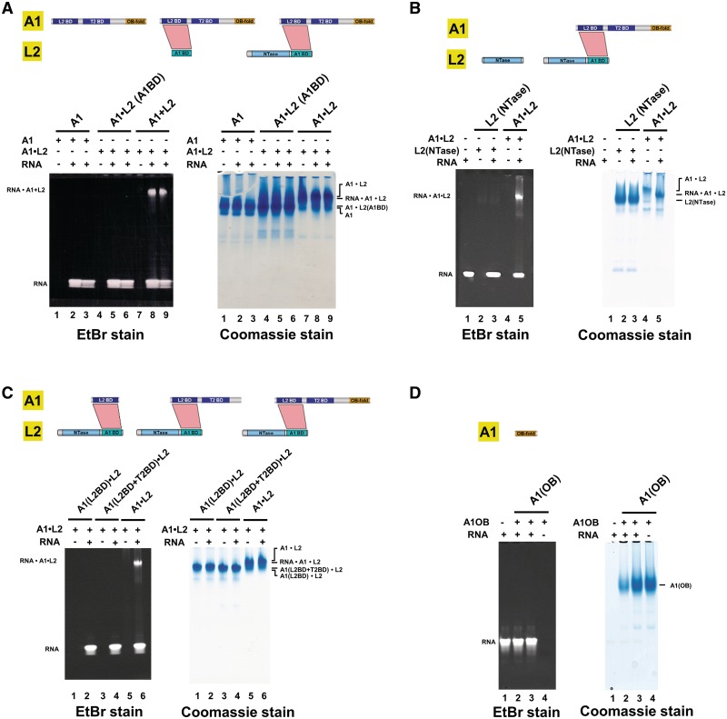
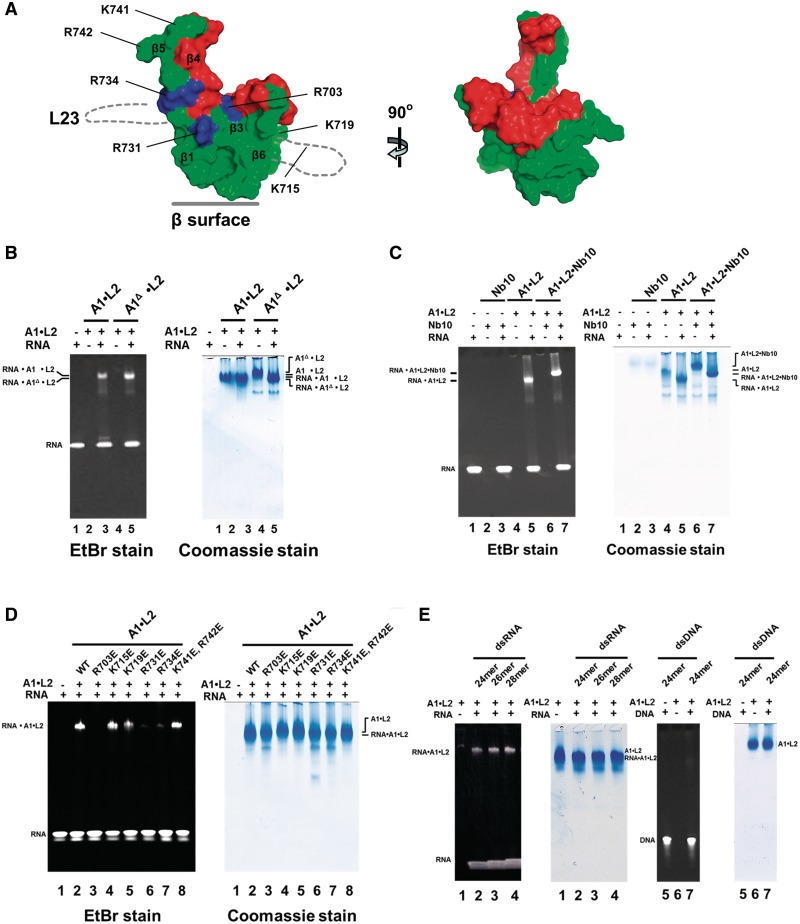
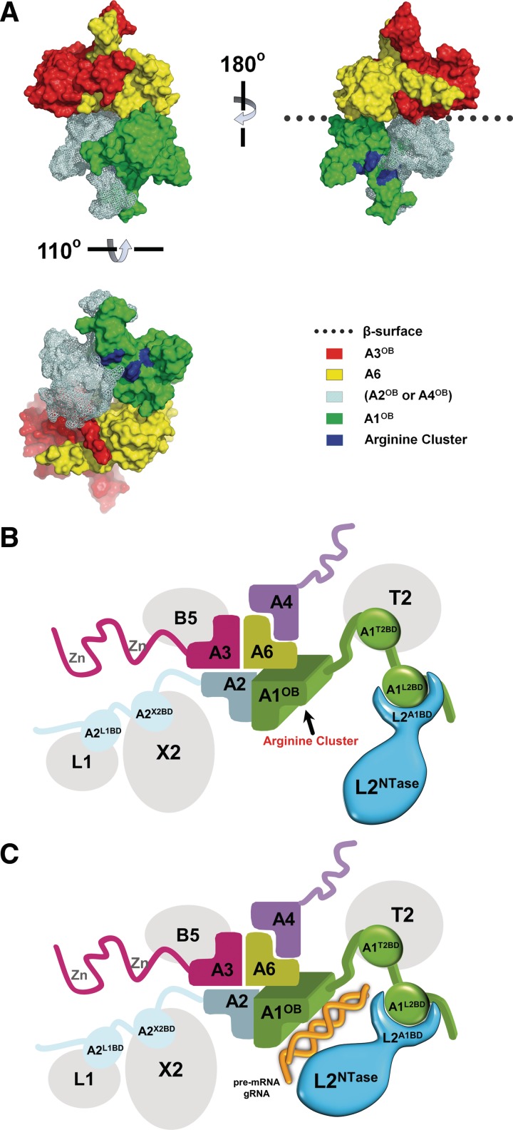
Trypanosomatids, such as the sleeping sickness parasite Trypanosoma brucei, contain a ∼ 20S RNA-editing complex, also called the editosome, which is required for U-insertion/deletion editing of mitochondrial mRNAs. The editosome contains a core of 12 proteins including the large interaction protein A1, the small interaction protein A6, and the editing RNA ligase L2. Using biochemical and structural data, we identified distinct domains of T. brucei A1 which specifically recognize A6 and L2. We provide evidence that an N-terminal domain of A1 interacts with the C-terminal domain of L2. The C-terminal domain of A1 appears to be required for the interaction with A6 and also plays a key role in RNA binding by the RNA-editing ligase L2 in trans. Three crystal structures of the C-terminal domain of A1 have been elucidated, each in complex with a nanobody as a crystallization chaperone. These structures permitted the identification of putative dsRNA recognition sites. Mutational analysis of conserved residues of the C-terminal domain identified Arg703, Arg731 and Arg734 as key requirements for RNA binding. The data show that the editing RNA ligase activity is modulated by a novel mechanism, i.e. by the trans-acting RNA binding C-terminal domain of A1.


Figures

Figure 1.
Figure 1.
T. brucei A1OB and A6 interact with each other. (A) The T2-binding domain (T2BD) and OB-fold domain arrangement in A1. A schematic representation of full length A1 and full length A6 with their interaction domains is shown with the T2BD of A1 in blue and the OB folds of A1 and A6 in gold. Direct interactions, as identified by bacterial co-expression and purification, are shown as a red polygon. The T2-binding domain (T2BD) of A1 had been identified before (17). (B) Co-transformation and expression of A1OB and A6 constructs. T. brucei A6 containing a His6-tag at the N-terminus co-transformed into Escherichia coli cells with A1OB (residues 626–762) was co-expressed. Cells were lysed and the His6-tagged A6•A1OB complex was captured by incubating the lysate with Ni-NTA beads (lane 2–4). The His6-tag peptide was cleaved off by TEV. This loss of the His6-tag peptide is evident in the reduced size of the now untagged A6 (lane 5). Abbreviations used in this and in the following figures: Cell, total lysate from induced cells; SF, soluble fraction; E, Ni-NTA elution fractions; E + TEV, eluate from first Ni-NTA after treatment with TEV protease; TEV, tobacco etch virus (TEV) protease. The positions and sizes (kDa) of marker polypeptides are indicated on the left.
Figure 2.
Figure 2.
The crystal structure of T. brucei A1OBΔ in complex with A1Nb10. (A) Overall structure of the A1OBΔA1Nb10 complex. The structure is shown as a ribbon diagram with the A1OBΔ domain and A1Nb10 depicted in green and gold, respectively. Selected secondary structure elements are labeled. The approximate location of the β-surface is indicated with a solid line. Two disordered regions are shown with dashed lines. (B) Electrostatic surface representations for A1OBΔ. The A1OBΔ domain is shown with its electrostatic surface charge and the nanobody A1Nb10 as a ribbon diagram. The electrostatic potential surface of A1OBΔ was calculated using APBS (51). Regions with potentials above +5 kbTec−1 and below 5 kbTec−1 are shown in blue and red, respectively. The locations of several conserved basic residues are indicated. (C) A side view of the complex shows that the basic residues on the surface of the A1OBΔ are facing away from nanobody binding site.
Figure 3.
Figure 3.
Identification of interaction domains of T. brucei A1 and L2. (A) A1L2BD and L2A1BD interact with each other. A schematic representation of the domains of full length A1 and full length L2, i.e. the L2-binding domain (L2BD), the T2-binding domain (T2BD), and the OB-fold domain (OB-Fold) of A1, and the nucleotidyl-transferase domain (NTase) and the A1-binding domain (A1BD) of L2. The direct interaction between the L2BD and the A1BD, as identified by bacterial co-expression and purification (see Figure 3B), is shown as a red polygon. (B) Co-transformation and co-expression of A1 L2BD and L2 A1BD constructs. T. brucei A1L2BD (residues 196–331) and L2A1BD (residues 308–416) were co-expressed in E. coli and co-purified by Ni-NTA chromatography via the His6-tag of L2 (lanes 2–4). The His6-tag of L2 was removed with TEV protease. This loss of the His6-tag of L2 is indicated by an arrow (lane 5). The positions and sizes (kDa) of marker polypeptides are indicated on the left (lane 1). See Figure 1B for abbreviations.
Figure 4.
Figure 4.
The C-terminal domain of T. brucei A1 promotes dsRNA binding by the RNA-editing ligase L2. Electrophoretic mobility shift assays were used to examine the binding of A1•L2 to nicked dsRNA substrate. Wild type and variants of A1•L2 binary complexes were co-expressed and co-purified as explained in Supplementary Methods. A total of 30 μM double-stranded 26-mer nicked RNA (see the sequence in Supplementary Table S2) was incubated with 15 μM proteins or protein complexes for 30 min at 4°C in 20 mM Tris (pH 7.5), 2 mM DTT and 250 mM NaCl. The RNA and A1•L2 complex were resolved on a 4–15% polyacrylamide gel and stained by ethidium bromide and Coomassie Blue, shown, respectively, at the left and the right of A–D. Each protein and the A1•L2 constructs used for the RNA-binding assays are illustrated in a schematic diagram above the gels. The positions of the RNA•A1•L2 complex and of RNA alone are indicated on the left. (A) The role of different domains of L2 in dsRNA recognition. The RNA binding abilities of (i) full length A1 (lanes 1–3) by itself, (ii) L2A1BD in complex with full length A1 (lanes 4–6) and (iii) full length L2(NTase+A1BD) with full length A1 (lanes 7–9), show that the NTase domain of L2 is critical for nicked dsRNA recognition. (B) The role of the L2NTase domain in dsRNA recognition. The RNA binding abilities of L2NTase domain by itself (lanes 2–3) and of wild-type full length A1 in complex with full length L2 (lanes 4–5), show that L2NTase alone fails to bind tightly to nicked dsRNA. Lane 1 contains 26-mer nicked dsRNA alone as a control. (C) The role of different domains of A1 in dsRNA recognition. The RNA binding abilities of (i) A1L2BD with full length L2 (lanes 1–2), (ii) A1(L2BD+T2BD) with full length L2 (lanes 3–4) and (iii) A1(L2BD+T2BD+OB) with full length L2 (lanes 5–6), show that the A1OB domain promotes RNA binding by L2. (D) The A1OB domain alone has little affinity for dsRNA. The RNA binding abilities of 15 and 45 μM A1OB alone (lanes 2–3) were tested. Lane 1 contains 26-mer nicked dsRNA alone as a control.
Figure 5.
Figure 5.
Identification of specific arginine residues involved in dsRNA binding by the T. brucei A1OB domain in complex with the RNA-editing ligase L2. (A) Surface presentation of the A1OBΔ structure. Left: the structure of A1OBΔ, with A1Nb10 omitted for clarity, is shown with (i) the residues involved in binding of A1Nb10 in red, (ii) the completely conserved and solvent exposed basic residues R703, R731 and R734 in blue and (iii) the remainder of the A1OBΔ surface in green. The approximate locations of the two disordered regions are sketched with dotted lines. Right: the A1OBΔ structure rotated ∼90° with respect to the left panel illustrating the A1Nb10-binding surface of A1OB which is not engaged in RNA-binding (Figure 5C). (B–E) Wild-type and variants of A1•L2 binary complexes were co-expressed and co-purified. Electrophoretic mobility shift assays—as explained in the Legend to Figure 4—were used to evaluate the RNA-binding capability of the set of A1•L2 variants obtained. (B) The effect of the deletion of L23 on RNA affinity of A1OB. The RNA binding abilities of wild-type A1•L2 (lanes 2 and 3) and A1Δ•L2 (lanes 4 and 5) are compared. Lane 1 contains 26-mer nicked dsRNA alone as a control. (C) The effect of A1Nb10 binding on RNA-binding activity. The RNA binding abilities of A1Nb10 alone (lanes 2 and 3), A1•L2 (lanes 4 and 5), and A1•L2• A1Nb10 (lanes 6 and 7) were compared. Lanes 1 contains 26-mer nicked dsRNA alone as a control. (D) Mutational effects on RNA-binding activity. The RNA-binding assay was carried out with single or double mutants of A1 with wild-type L2. Each of seven basic residues (R703, K715, K719, R731, R734, K741 and R742) in full length A1 was replaced by glutamate as indicated (lanes 3–8). The RNA binding abilities of wild-type and mutant A1•L2 were tested. 26-mer nicked dsRNA alone (lane 1) and wild-type A1•L2 (lane 2) were used as a control. (E) The preference for dsRNA over dsDNA substrates on RNA-binding activity. The binding of both nicked RNA duplex substrates and nicked DNA duplex substrates by recombinant A1•L2 was tested. To probe the possibility of sequence specificity on dsDNA recognition, two different random sequence dsDNA were also tested as a control and appeared not to bind to A1•L2 (data not shown).
Figure 6.
Figure 6.
Model of the OB-fold center and associated proteins in the core of the editosome. (A) A ‘shifted heterotetramer’ model of the OB folds of A1, A2 or A4, A3 and A6. The shifted heterotetramer described in (29) was modified by replacing the model structure for A1OB by the actual structure of A1OB described in this article. The heterotetramer depicted contains the actual structure of the A3OB•A6 heterodimer (29), the new structure of A1OB (Figure 5A and Supplementary Figure S4) and a model of A2OB based on the structure of A1OB. The A2OB domain might be an A4OB domain instead, as discussed in (29). (B) The ‘shifted heterotetramer’ model with four associated enzymes depicted schematically. The OB domains are colored as in (A); the editing RNA ligase L2 is shown in blue; four additional editosome proteins in gray. The three domains of A1 are schematically shown interacting with partner proteins in the core: the A1L2BD with the L2A1BD, A1T2BD domain with T2 and A1OB with A6. The editosome proteins schematically depicted are: TUTase 2 (T2), Exonuclease 2 (X2), Ligase 1 (L1), Ligase 2 (L2), interaction proteins A1 to A6 (A1–A6) and KREPB5 (B5). Interaction data is from references (15,17) and from Figures 1, 3 and 4. (C). A1 and L2 recognizing dsRNA. Color code as in (B) above with dsRNA schematically added in gold, and with the L2NTase domain closer to A1OB. The A1L2BD and L2A1BD domains interact with each other, thereby enabling the A1OB and L2NTase domains to cooperatively bind dsRNA substrate. For further explanation see ‘Discussion’ section.

References

    1. Schnaufer A, Domingo GJ, Stuart K. Natural and induced dyskinetoplastic trypanosomatids: how to live without mitochondrial DNA. Int. J. Parasitol. 2002;32:1071–1084. - PubMed
    1. Vickerman K. Polymorphism and mitochondrial activity in sleeping sickness trypanosomes. Nature. 1965;208:762–766. - PubMed
    1. Koslowsky DJ, Riley GR, Feagin JE, Stuart K. Guide RNAs for transcripts with developmentally regulated RNA editing are present in both life cycle stages of Trypanosoma brucei. Mol. Cell Biol. 1992;12:2043–2049. - PMC - PubMed
    1. Stuart K. RNA editing in trypanosomatid mitochondria. Annu. Rev. Microbiol. 1991;45:327–344. - PubMed
    1. Aphasizhev R, Aphasizheva I. Mitochondrial RNA processing in trypanosomes. Res. Microbiol. 2011;162:655–663. - PMC - PubMed

Publication types

MeSH terms