

Differential effects of m1 and m2 receptor antagonists in perirhinal cortex on visual recognition memory in monkeys
- PMID: 22561485
- PMCID: PMC3389587
- DOI: 10.1016/j.nlm.2012.04.007
Differential effects of m1 and m2 receptor antagonists in perirhinal cortex on visual recognition memory in monkeys
Abstract
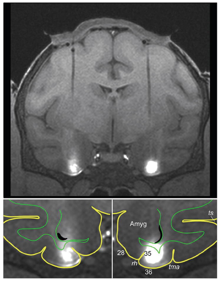
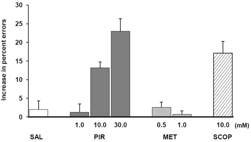
Microinfusions of the nonselective muscarinic antagonist scopolamine into perirhinal cortex impairs performance on visual recognition tasks, indicating that muscarinic receptors in this region play a pivotal role in recognition memory. To assess the mnemonic effects of selective blockade in perirhinal cortex of muscarinic receptor subtypes, we locally infused either the m1-selective antagonist pirenzepine or the m2-selective antagonist methoctramine in animals performing one-trial visual recognition, and compared these scores with those following infusions of equivalent volumes of saline. Compared to these control infusions, injections of pirenzepine, but not of methoctramine, significantly impaired recognition accuracy. Further, similar doses of scopolamine and pirenzepine yielded similar deficits, suggesting that the deficits obtained earlier with scopolamine were due mainly, if not exclusively, to blockade of m1 receptors. The present findings indicate that m1 and m2 receptors have functionally dissociable roles, and that the formation of new visual memories is critically dependent on the cholinergic activation of m1 receptors located on perirhinal cells.
Published by Elsevier Inc.
Conflict of interest statement
The authors declare they have no competing financial interests.
Figures
References
-
- Aigner TG, Mishkin M. The effects of physostigmine and scopolamine on recognition memory in monkeys. Behav Neural Biol. 1986;45(1):81–87. - PubMed
-
- Aigner TG, Walker DL, Mishkin M. Comparison of the effects of scopolamine administered before and after acquisition in a test of visual recognition memory in monkeys. Behav Neural Biol. 1991;55(1):61–67. - PubMed
-
- Alcantara AA, Mrzljak L, Jakab RL, Levey AI, Hersch SM, Goldman-Rakic PS. Muscarinic m1 and m2 receptor proteins in local circuit and projection neurons of the primate striatum: anatomical evidence for cholinergic modulation of glutamatergic prefronto-striatal pathways. J Comp Neurol. 2001;434(4):445–460. - PubMed
-
- Birdsall NJ, Curtis CA, Eveleigh P, Hulme EC, Pedder EK, Poyner D, et al. Muscarinic receptor subtypes and the selectivity of agonists and antagonists. Pharmacology. 1988;37(Suppl 1):22–31. - PubMed
Publication types
MeSH terms
Substances
Grants and funding
LinkOut - more resources
Full Text Sources
