Review
. 2013 Jan;1834(1):443-53.
doi: 10.1016/j.bbapap.2012.04.004. Epub 2012 Apr 26.

Catalytic mechanisms for phosphotriesterases

Affiliations
Review

Catalytic mechanisms for phosphotriesterases

Andrew N Bigley et al. Biochim Biophys Acta. 2013 Jan.

Abstract

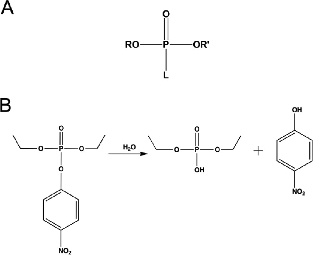
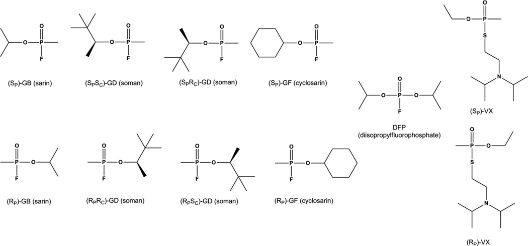
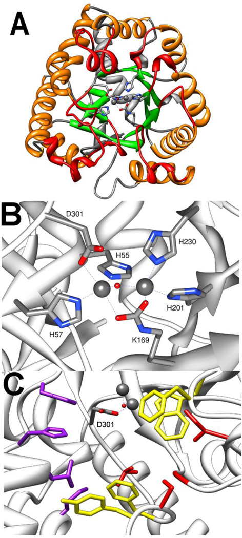
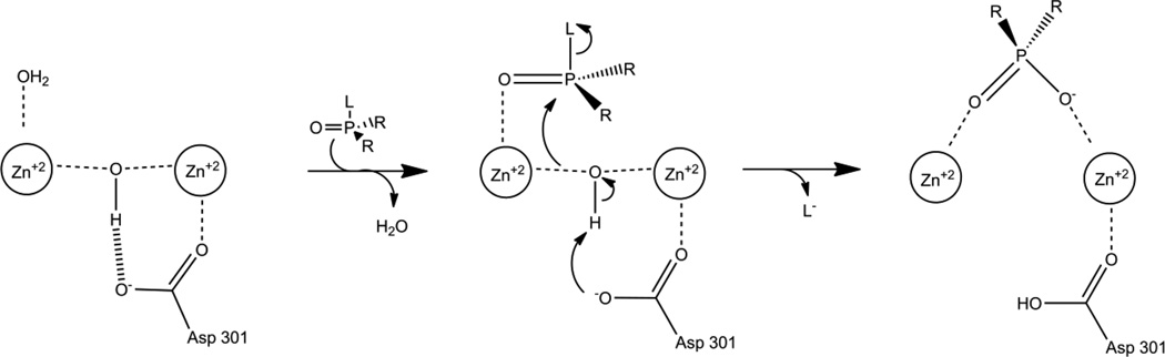
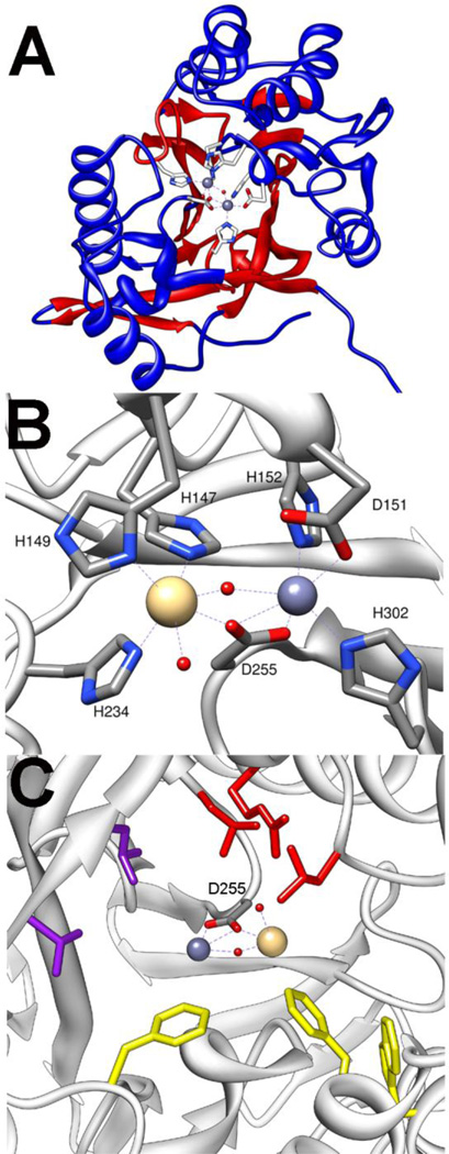
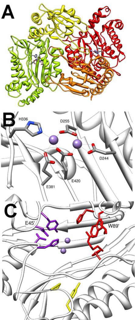
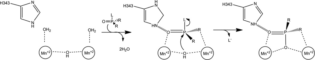
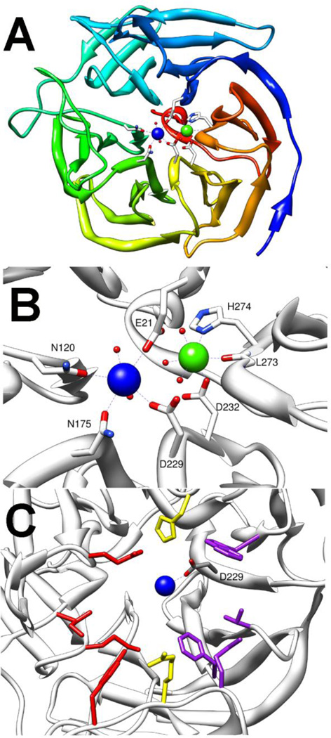
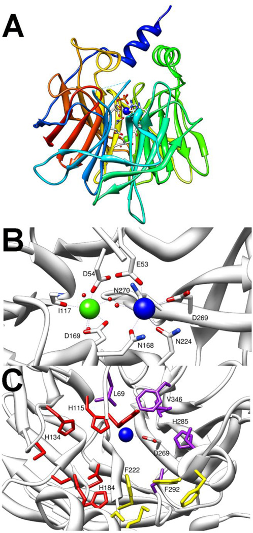
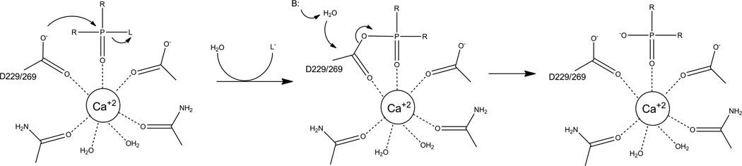
Phosphotriesters are one class of highly toxic synthetic compounds known as organophosphates. Wide spread usage of organophosphates as insecticides as well as nerve agents has lead to numerous efforts to identify enzymes capable of detoxifying them. A wide array of enzymes has been found to have phosphotriesterase activity including phosphotriesterase (PTE), methyl parathion hydrolase (MPH), organophosphorus acid anhydrolase (OPAA), diisopropylfluorophosphatase (DFP), and paraoxonase 1 (PON1). These enzymes differ widely in protein sequence and three-dimensional structure, as well as in catalytic mechanism, but they also share several common features. All of the enzymes identified as phosphotriesterases are metal-dependent hydrolases that contain a hydrophobic active site with three discrete binding pockets to accommodate the substrate ester groups. Activation of the substrate phosphorus center is achieved by a direct interaction between the phosphoryl oxygen and a divalent metal in the active site. The mechanistic details of the hydrolytic reaction differ among the various enzymes with both direct attack of a hydroxide as well as covalent catalysis being found. This article is part of a Special Issue entitled: Chemistry and mechanism of phosphatases, diesterases and triesterases.


Figures

Figure 1
Figure 1
A) General structure of an organophosphate where R and R' are alkyl groups and L can be a phenol, thiol, or fluoride group. B) Hydrolysis reaction of paraoxon catalyzed by phosphotriesterase.
Figure 2
Figure 2
Chemical structures of organophosphate insecticides. Common examples are shown for A) Phosphotriesters, B) Thiophosphates, C) Phosphorothiolates.
Figure 3
Figure 3
Chemical structures for the individual enantiomers of chiral organophosphate nerve agents GB (sarin), GD (soman), GF (cyclosarin) and VX as well as the achiral analog DFP. Chiral phosphorus centers are drawn as Fischer projections. The Additional stereo-center in GD is drawn in standard stereochemical convention.
Figure 4
Figure 4
Crystal structure of PTE (pdb. 1dpm). A) TIM-Barrel fold of PTE shown with core α-helices in orange, core β-strands shown in green, N-terminal loops shown in grey, and C-terminal loops shown in red. Metals in active site are shown as spheres, and the metal ligating side chains shown as sticks. B) Metal binding site of PTE. C) Substrate binding pockets of PTE. Leaving group pocket residues W131, F132, F306, and Y309 are shown in yellow. Large group pocket residues H254, H257, L271, M317 are shown in purple. Small group pocket residues G60, I106, L303, and S308 are shown in red.
Figure 5
Figure 5
Chemical structures of enantiomers of chiral phosphate and phosphonate nerve agent analogs for GB and GF. Chiral phosphorus centers are drawn as Fischer projections.
Figure 6
Figure 6
Proposed catalytic mechanism of PTE.
Figure 7
Figure 7
Crystal structure of MPH (pdb. 1p9e). A) αβ/βα structure of monomer shown with central β-strands in red and the sandwiching α-helices shown in blue. Metals at active site are shown as spheres and ligating residues shown as sticks. B) Metal site shown with the smaller Zn2+ in α-site and the larger Cd2+ in the β-site. C) Substrate binding pockets of MPH. Leaving group pocket residues are shown in yellow. Side pockets are shown in purple and red.
Figure 8
Figure 8
Crystal structure of OPAA (pdb. 3l7g). A) Dimeric structure of OPAA. One protomer is shown with N-terminal domain in yellow and C-terminal domain green. The second protomer is shown with N-terminal domain in orange and C-terminal domain in red. Mn2+ is shown as spheres, and metal ligating residues are shown as sticks. B) Expanded view of the OPAA metal center. C) Substrate binding site of OPAA. Small binding pocket residues are shown in purple. Large binding pocket residues are colored red. Leaving group pocket is colored yellow. The residues in large and small pockets from opposing subunit are labeled.
Figure 9
Figure 9
Proposed catalytic mechanism for OPAA cleavage of phosphotriesters. L is leaving group which can be fluoride, or p-nitrophenol. R is an ester or methyl group.
Figure 10
Figure 10
Crystal structure of DFPase (pdb 2gvv). Catalytic calcium is shown in blue while structural calcium is shown in green. A) Top down view of DFPase showing the 6 bladed β-propeller structure. B) Metal center ligation of DFPase. C) Substrate binding site in DFPase. Side pocket residues Y144, M90, I72 and E37 are shown in red. In purple are W244, T195, F173, and M148 from the second side pocket. The central cleft residues R146 and H287 are shown in yellow. Catalytic aspartate is labeled.
Figure 11
Figure 11
Crystal structure of PON1 (pdb. 1v04). A) Side view of β-propeller fold with HDL anchoring helices extending above. Catalytic calcium shown in blue, structural calcium shown in green, and ligating residues shown as sticks. B) Metal centers and ligating residues of PON1. Catalytic calcium is blue structural calcium is green. C) Substrate binding pockets of PON1. Large group pocket residues are colored red. Small group pocket is colored purple, and leaving group pocket is colored yellow. Catalytic aspartate (D269) and residues known to be important for the phosphotriesterase reaction are labeled.
Figure 12
Figure 12
Proposed catalytic mechanism for phosphotriesterase reaction catalyzed by PON1 and DFPase. R is ester linked alcohol group or a methyl group. L is leaving group which is fluoride for DFPase or for PON1 a fluoride, a phenol, or a thiol.

References

    1. Raushel FM. Bacterial detoxification of organophosphate nerve agents. Curr. Opin. Microbiol. 2002;5:288–295. - PubMed
    1. Musilek K, Dolezal M, Gunn-Moore F, Kuca K. Design, evaluation and structure---Activity relationship studies of the AChE reactivators against organophosphorus pesticides. Med. Res Rev. 2011;31:548–575. - PubMed
    1. Munro NB, Ambrose KR, Watson AP. Toxicity of the organophosphate chemical warfare agents GA, GB, and VX: Implications for public protection. Environ. Health Perspect. 1994;102:18–38. - PMC - PubMed
    1. Harper LL, McDaniel CS, Miller CE, Wild JR. Dissimilar plasmids isolated from Pseudomonas diminuta MG and a Flavobacterium sp. (ATCC 27551) contain identical opd genes. Appl. Environ. Microbiol. 1988;54:2586–2589. - PMC - PubMed
    1. Sun L, Dong Y, Zhou Y, Yang M, Zhang C, Rao Z, Zhang X. Crystalization and preliminary X-ray studies of methyl parathion hydrolase from Pseudomonas sp. WBC-3. Acta Crystallogr. D Biol. Crystallogr. 2004;60:954–956. - PubMed