. 2012 Jun;135(Pt 6):1819-33.
doi: 10.1093/brain/aws105. Epub 2012 May 4.

Functional identification of pathogenic autoantibody responses in patients with multiple sclerosis

Affiliations

Functional identification of pathogenic autoantibody responses in patients with multiple sclerosis

Christina Elliott et al. Brain. 2012 Jun.

Abstract

Pathological and clinical studies implicate antibody-dependent mechanisms in the immunopathogenesis of multiple sclerosis. We tested this hypothesis directly by investigating the ability of patient-derived immunoglobulins to mediate demyelination and axonal injury in vitro. Using a myelinating culture system, we developed a sensitive and reproducible bioassay to detect and quantify these effects and applied this to investigate the pathogenic potential of immunoglobulin G preparations obtained from patients with multiple sclerosis (n = 37), other neurological diseases (n = 10) and healthy control donors (n = 13). This identified complement-dependent demyelinating immunoglobulin G responses in approximately 30% of patients with multiple sclerosis, which in two cases was accompanied by significant complement-dependent antibody mediated axonal loss. No pathogenic immunoglobulin G responses were detected in patients with other neurological disease or healthy controls, indicating that the presence of these demyelinating/axopathic autoantibodies is specific for a subset of patients with multiple sclerosis. Immunofluorescence microscopy revealed immunoglobulin G preparations with demyelinating activity contained antibodies that specifically decorated the surface of myelinating oligodendrocytes and their contiguous myelin sheaths. No other binding was observed indicating that the response is restricted to autoantigens expressed by terminally differentiated myelinating oligodendrocytes. In conclusion, our study identifies axopathic and/or demyelinating autoantibody responses in a subset of patients with multiple sclerosis. This observation underlines the mechanistic heterogeneity of multiple sclerosis and provides a rational explanation why some patients benefit from antibody depleting treatments.


Figures

Figure 1
Figure 1
Myelinating cultures reproduce the major characteristics of in vivo CNS-myelinated axons. [A(I)] In vitro myelinating cultures consist of a network of SMI-31+ axons (red, phosphorylated neurofilament staining) some of which are myelinated by PLP+ oligodendrocytes (green; ×20 magnification; scale bar = 100 µm). These myelin internodes are interspaced by nodes of Ranvier characterized by axonal expression of Nfasc186/NaV bounded by Caspr/Nfasc155 at the paranode. [A(II)] Caspr, blue; PLP, red; Nfasc186, green (×100 magnification, scale bar = 10 µm); [A(III)] Nfasc155, blue; PLP, green; NaV, red (×100 magnification, scale bar = 10 µm). (B) To validate using these myelinating cultures as a target for pathogenic antibody responses, we used a panel of antibodies against a number of targets previously implicated in multiple sclerosis. In each case, cultures were treated overnight with 10 µg/ml antibody in the presence of 1% fresh rat serum as a source of complement. Axonal density and myelination were quantified using computer aided analysis of immunofluorescent images. The pathogenic activity of each antibody was highly selective and dependent on its targets localization. Treatment with antibodies recognizing antigens accessible at the myelin/oligodendrocyte surface such as MOG (Z2/8-18C5) or sulphatide (O4) results in complete and selective demyelination with no significant axonal injury. In contrast, antibodies recognizing intracellular epitopes such as myelin basic protein (MBP) or the C-terminus of PLP were unable to induce myelin loss. Targeting axons using an antibody against Nfasc186 mediates significant axonal loss and secondary demyelination. These antibody-mediated effects were complement-dependent as heat inactivation of serum abolished all antibody mediated effects. Antibody activity is also antigen specific as demonstrated using isotype controls (MOPC1, UPC10). Percentage values for axonal loss and demyelination were calculated in relation to untreated cultures (mean ± SD, n = 3; *P < 0.05, **P < 0.001, t-test).
Figure 2
Figure 2
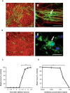
Targeting MOG induces selective demyelination without causing axonal injury. [A(I)] Overnight treatment of myelinating cultures with 10 µg/ml Z2 (MOG-specific monoclonal antibody) in the absence of complement does not induce axonal injury as visualized by intact SMI-31 (phosphorylated neurofilament) staining (red) or demyelination visualized with PLP (green; ×10 magnification; scale bar = 100 µm). High magnification images show oligodendrocytes are unaffected after overnight treatment with Z2 in the absence of a source of complement [A(II)]; ×60 magnification; scale bar = 50 µm). [B(I)] In contrast, antibody treatment in the presence of fresh rat sera as a source of complement (1%)-induced complete demyelination and loss of oligodendrocytes but leaving an intact bed of axons (×10 magnification; scale bar = 100 µm). [B(II)] Antibody-mediated demyelination is associated with oligodendrocyte lysis (PLP, green; nucleus, blue; ×100 magnification; scale bar = 50 µm). (C) Demyelination occurs rapidly after antibody addition. Significant demyelination was first detected after 3.5 h and by 4 h demyelination is virtually complete. Percentage values for axonal loss and demyelination were calculated in relation to cultures treated with equivalent antibody concentrations in the presence of heat inactivated complement (mean ± SD, n = 3; **P < 0.001, t-test). (D) Myelinating cultures provide a highly sensitive screening strategy in which to detect pathogenic autoantibody responses. Significant demyelinating activity is detectable at antibody concentrations ≥50 ng/ml (∼300 pM). Percentage values of demyelination were calculated in relation to cultures treated with equivalent concentrations of an isotype control antibody (mean ± SD, n = 3; **P < 0.001, t-test).
Figure 3
Figure 3
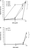
Dose dependence of multiple sclerosis derived autoantibody-mediated pathogenesis. IgG from four patients with multiple sclerosis (Patients MS2, MS4, MS5 and MS14) known to contain axopathic and/or demyelinating activity was added to myelinating cultures at 100, 50, 10 and 1 µg/ml in the presence of 1% fresh rat serum as a source of complement. (A) Demyelinating activity in all cases was significantly reduced at 50 µg/ml and was no longer detected at 1 µg/ml. (B) Axopathic activity present in samples from Patients MS5 and MS14 was not detected at IgG concentrations <100 µg/ml. Percentage values for axonal loss and demyelination were calculated in relation to cultures treated with equivalent antibody concentrations in the presence of heat inactivated complement (mean ± SD, n = 3; **P < 0.001, t-test). Ab = antibody.
Figure 4
Figure 4
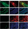
Patient derived IgG preparations with demyelinating activity decorate the surface of myelin sheaths and contiguous oligodendrocytes. [A(IIII)] Live staining of myelinating cultures with patient derived IgG preparations with in vitro demyelinating activity revealed binding to surface of PLP+ myelin sheaths and oligodendrocytes (human IgG green; PLP, red) and align with SMI-31+ axons [A(IVVI)] (human IgG, green; SMI-31, blue; ×60 magnification; scale bar = 20 µm). [B(IIII)] Live staining of myelinating cultures with human IgG preparations with no axopathic or demyelinating activity fails to identify any specific recognition of PLP+ myelin sheaths and oligodendrocytes (human IgG green; PLP, red) or [B(IVVI)] SMI-31+ axons (human IgG, green; SMI-31, blue; ×60 magnification; scale bar = 20 µm).
Figure 5
Figure 5
The autoantigen(s) recognized by pathogenic multiple sclerosis IgG are expressed by mature and myelinating oligodendrocytes. Schematic representation of the oligodendrocyte lineage differentiation pathway. NG2 is only expressed by oligodendrocyte progenitor cells (OPC) early in differentiation. Sulphatide (O4) is expressed later by oligodendrocyte progenitor cells, pre-oligodendrocytes, mature oligodendrocytes (OLG) and myelin. PLP is expressed on mature oligodendrocytes and myelin, however, it must be noted that DM20, an isoform of PLP, is expressed earlier by a population of pre-oligodendrocytes. From our data, we can determine that the pathogenic antibodies detected in multiple sclerosis IgG are directed against antigens expressed on mature oligodendrocytes and myelin as antibody treatment spared the majority of NG2+ and O4+ cells. The shaded area represents the relative proportion of cells lost in responses to treatment with multiple sclerosis patient derived IgG.

References

    1. Appel SH, Bornstein MB. The application of tissue culture to the study of experimental allergic encephalomyelitis. II Serum factors responsible for demyelination. J Exp Med. 1964;119:303–12. - PMC - PubMed
    1. Archelos JJ, Hartung HP. Pathogenetic role of autoantibodies in neurological diseases. Trends Neurosci. 2000;23:317–27. - PubMed
    1. Barnett MH, Parratt JD, Cho ES, Prineas JW. Immunoglobulins and complement in postmortem multiple sclerosis tissue. Ann Neurol. 2009;65:32–46. - PubMed
    1. Bornstein MB, Murray MR. Serial observations on patterns of growth, myelin formation, maintenance and degeneration in cultures of newborn rat and kitten cerebellum. J Biophys Biochem Cytol. 1958;4:499–504. - PMC - PubMed
    1. Bradl M, Misu T, Takahashi T, Watanabe M, Mader S, Reindl M, et al. Neuromyelitis optica: pathogenicity of patient immunoglobulin in vivo. Ann Neurol. 2009;66:630–43. - PubMed

Publication types

MeSH terms