Review
. 2012 May 4;18(5):705-15.
doi: 10.1038/nm.2760.

T cell homing to epithelial barriers in allergic disease

Affiliations
Review

T cell homing to epithelial barriers in allergic disease

Sabina A Islam et al. Nat Med. .

Abstract

Allergic inflammation develops in tissues that have large epithelial surface areas that are exposed to the environment, such as the lung, skin and gut. In the steady state, antigen-experienced memory T cells patrol these peripheral tissues to facilitate swift immune responses against invading pathogens. In at least two allergy-prone organs, the skin and the gut, memory T cells are programmed during the initial antigen priming to express trafficking receptors that enable them to preferentially home to these organs. In this review we propose that tissue-specific memory and inflammation-specific T cell trafficking facilitates the development of allergic disease in these organs. We thus review recent advances in our understanding of tissue-specific T cell trafficking and how regulation of T cell trafficking by the chemokine system contributes to allergic inflammation in mouse models and in human allergic diseases of the skin, lung and gut. Inflammation- and tissue-specific T lymphocyte trafficking pathways are currently being targeted as new treatments for non-allergic inflammatory diseases and may yield effective new therapeutics for allergic diseases.


Figures

Figure 1
Figure 1
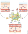
Generation of allergen-specific effector and memory T cells during primary mmune responses at the epithelial barrier. During the primary immune response to allergens, disruption of the epithelial barrier eads to epithelial cell production of cytokines, such as TSLP, IL-25 and IL-33. Allergens and other inflammatory cues in the presence of TSLP, IL-25 and IL-33 induce dendritic cell (DC) maturation and trafficking to tissue draining lymph nodes (LN) where dendritic cells activate naive T (TN) cells to differentiate into TH2-type effector (TEFF) cells. Allergen-specific TEFF cells undergo clonal proliferation in the lymph nodes, and TEFF cells exit the ymph nodes through efferent lymphatics. TEFF cells re-enter the systemic circulation through the thoracic duct and enter inflamed tissue through postcapillary venules. The majority of TEFF cells eventually die, but a minority differentiate into memory T (TM) cells several weeks after the resolution of nflammation. Depending on the profile of trafficking receptors expressed, different subsets of TM cells perform distinct types of mmune surveillance in the steady state. Naive T cells and central memory T (TCM) cells equipped with L-selectin (CD62L) and the chemokine receptor CCR7 have restricted and specific access to secondary ymphoid tissue through specialized high endothelial venules (HEVs), which express their respective ligands (PNAd and CCL21). T cells recirculate between lymphoid tissue and blood. Effector memory T (TEM) cells primarily recirculate between the blood and peripheral tissue. TRM cells persist in peripheral tissue and do not recirculate. TSLPR, TSLP receptor; IL-33R, IL-33 receptor.
Figure 2
Figure 2
Tissue-specific imprinting and steady-state programmed memory T cell trafficking to atopy-prone organs. Dendritic cells of the skin and the gut imprint T cells during the primary immune response to express trafficking receptors that enable tissue-specific homing. Skin T cells express CLA, CCR10, CCR4 and CCR8, which enable skin TEM cell entry into the skin in the steady state, as postcapillary venules in the skin express CCL17, E-selectin and CCL1 (the respective igands for CCR4, CLA and CCR8) and epidermal cells expresses CCL27 (the ligand for CCR10) and mCCL8 (the ligand for CCR8 in the mouse). Gut TEM cells are programmed to express CCR9 and α4β7, which enable the entry of gut TEM cells into the small intestine through postcapillary venules that express CCL25 and MADCAM1, which are the respective ligands for CCR9 and α4β7. Skin- and gut-specific programmed lymphocyte homing pathways are well characterized in human and mouse studies. Human studies also suggest that programmed tissue-specific homing of lung TEM cells may also occur.
Figure 3
Figure 3
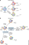
Chemokine receptors associated with CD4+ helper T cell subsets. (a) During the primary immune response, cytokines (indicated above the arrow) and transcription factors (T-bet, GATA3 and ROR-γT) nstruct naive T cells to differentiate into TH cell subsets. After TCR activation, specific cytokines derived from the innate immune system and unique master transcription factors direct naive CD4 TH cells to differentiate into TH1, TH2, TH17, Treg and TFH cell subsets. Differentiated TH cell subsets produce lineage-specific effector cytokines and express characteristic chemokine receptors. (b) After recall stimulation with an antigen, allergen-specific memory TH2 cells rapidly produce higher amounts of effector cytokines, such as IL-4, on rechallenge with the cognate allergen as compared to naive T cells. Repeated allergen-induced TH2 cell differentiation induces a highly differentiated, CCR8+IL-5hi TH2 cell subset that coexpresses the indicated receptors. Memory TH2 cells may not be fixed in differentiation along the TH2 lineage and may adopt features of TH17 and TH1 cells on rechallenge with allergen in the context of inflammatory cues derived from fungi or viruses, respectively. TGF-β, transforming growth factor β; IL-25R, IL-25 receptor.
Figure 4
Figure 4
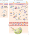
Trafficking of TH2 cell subsets into the skin during the acute and chronic phases of atopic dermatitis. During the acute phase of atopic dermatitis, the initiation of inflammation is dependent on TH2 cell entry into inflamed skin by CCR4 and the CCR4 ligand CCL17, which is upregulated in inflamed postcapillary venules. During the chronic phase of atopic dermatitis, the pool of allergen-specific skin tissue TRM cells is increased as compared to in the acute phase, and these allergen-specific TRM cells can initiate inflammation independent of CCR4-dependent TH2 cell entry from the blood. Repeated prior allergen exposure also leads to the in vivo generation of the more differentiated CCR8+IL-5hi TH2 cell subset in the chronic phase. Allergen challenge induces production of the CCR8 ligand, mCCL8, in the skin. mCCL8-dependent trafficking of L-5hi CCR8+ TH2 cells into the skin drives eosinophilic skin inflammation in the chronic phase of atopic dermatitis in the mouse model.
Figure 5
Figure 5
Chemokine-regulated TH cell trafficking into the lung during the early and late phases of the asthmatic response. During the early phase allergic airway response (left), allergen-IgE complexes stimulate airway mast cells to degranulate and release acute mediators such as histamine, tumor necrosis factor α (TNF-α), LTB4, PGD2 and the chemokine CCL1. LTB4, PGD2 and CCL1 recruit TH2 cells into the airways through BLT1, CRTH2 and CCR8, respectively. T lymphocyte trafficking during the late phase allergic airway response (right) is facilitated by innate cells, such as macrophages and myeloid dendritic cells (mDCs). Allergen-induced TLR activation induces macrophage and epithelial cell release of chemokines such as CXCL10 and CCL20. These chemokines recruit an initial wave of T cells into the airways through their receptors, CXCR3 and CCR6. IL-4 and IL-13 produced by activated lung allergen-specific TRM cells stimulate mDCs to release the chemokines CCL17 and CCL22 in a STAT6-dependent fashion. These chemokines amplify the subsequent recruitment of TH2 effector cells, which are generated in lung draining lymph nodes, into the airways from the circulation by the TH2 cell receptor CCR4. IL-4 and IL-13 also induce the differentiation of alternatively activated macrophages, which produce CCL17 to promote CCR4-dependent TH2 cell recruitment. CCR8 and CX3CR1 are also probably involved in T cell recruitment into the lung during the late phase allergic airway response. CCR8 and CX3CR1 are implicated in human asthma and in mouse models of airway inflammation. The CCR8 ligand mCCL8 is upregulated in mouse models of allergic airway inflammation, but the cellular sources of mCCL8 in the lung have not been defined. The CX3CR1 ligand, CX3CL1, is induced in airway smooth muscle cells, lung endothelium and epithelial cells after allergen challenge.

References

    1. Cookson W. The immunogenetics of asthma and eczema: a new focus on the epithelium. Nat. Rev. Immunol. 2004;4:978–988. - PubMed
    1. Rodriguez E, et al. Meta-analysis of filaggrin polymorphisms in eczema and asthma: robust risk factors in atopic disease. J. Allergy Clin. Immunol. 2009;123:1361–1370. - PubMed
    1. Al-Shami A, et al. A role for TSLP in the development of inflammation in an asthma model. J. Exp. Med. 2005;202:829–839. - PMC - PubMed
    1. Eiwegger T, Akdis CA. IL-33 links tissue cells, dendritic cells and Th2 cell development in a mouse model of asthma. Eur. J. Immunol. 2011;41:1535–1538. - PubMed
    1. Fort MM, et al. IL-25 induces IL-4, IL-5, and IL-13 and Th2-associated pathologiesin vivo . Immunity. 2001;15:985–995. - PubMed

Publication types