

Continuum modeling of a neuronal cell under blast loading
- PMID: 22562014
- PMCID: PMC3408831
- DOI: 10.1016/j.actbio.2012.04.039
Continuum modeling of a neuronal cell under blast loading
Abstract
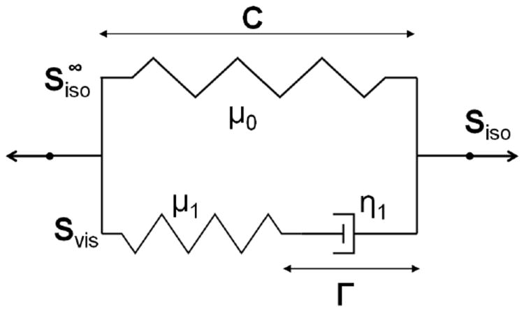
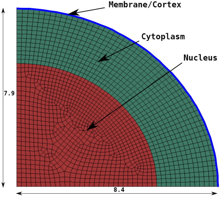
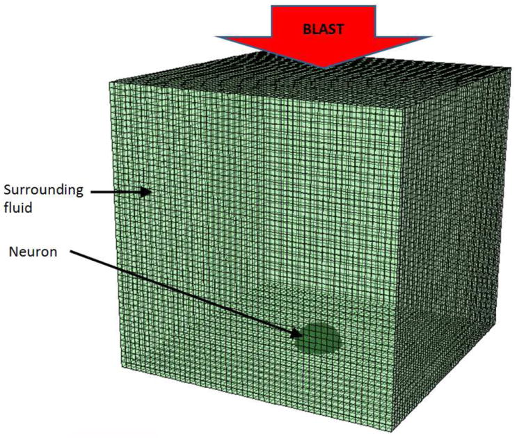
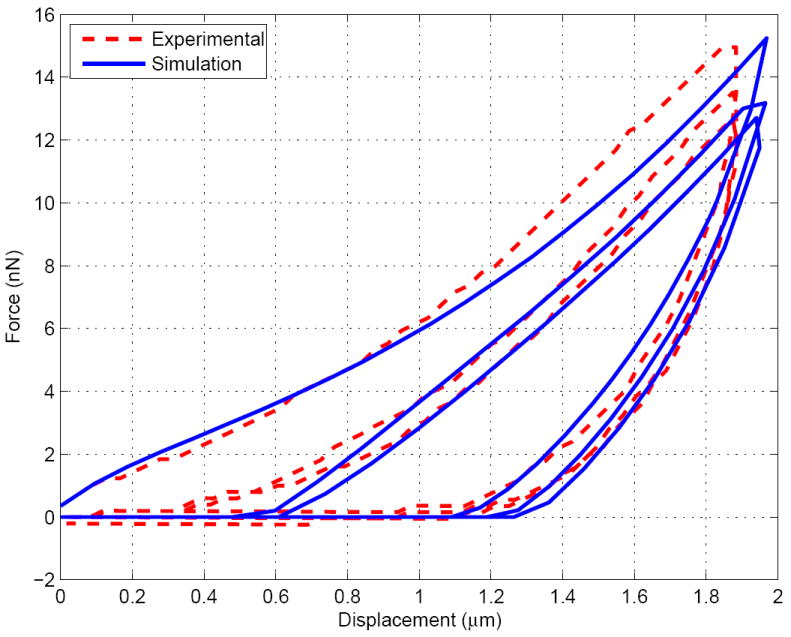
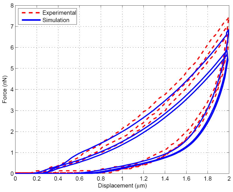
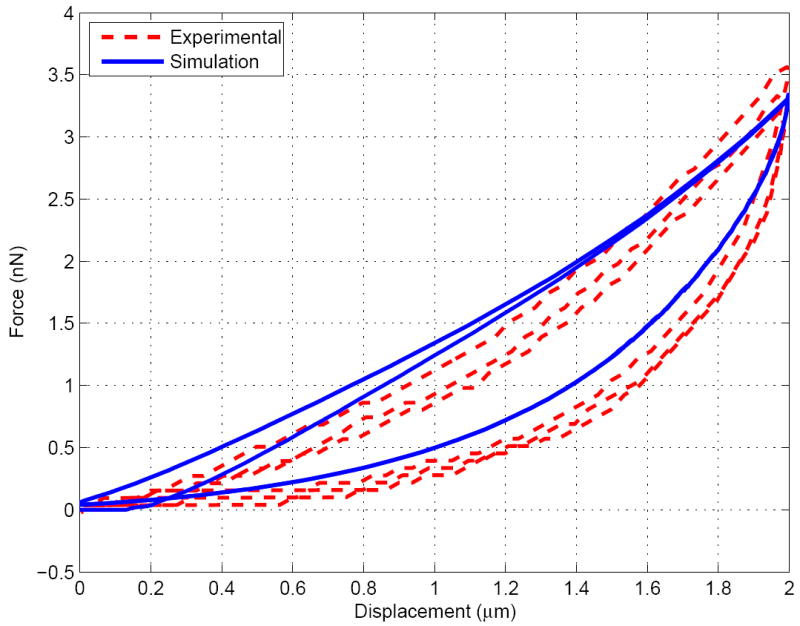
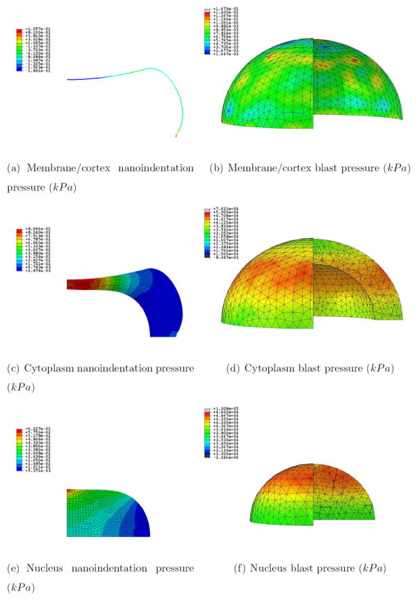
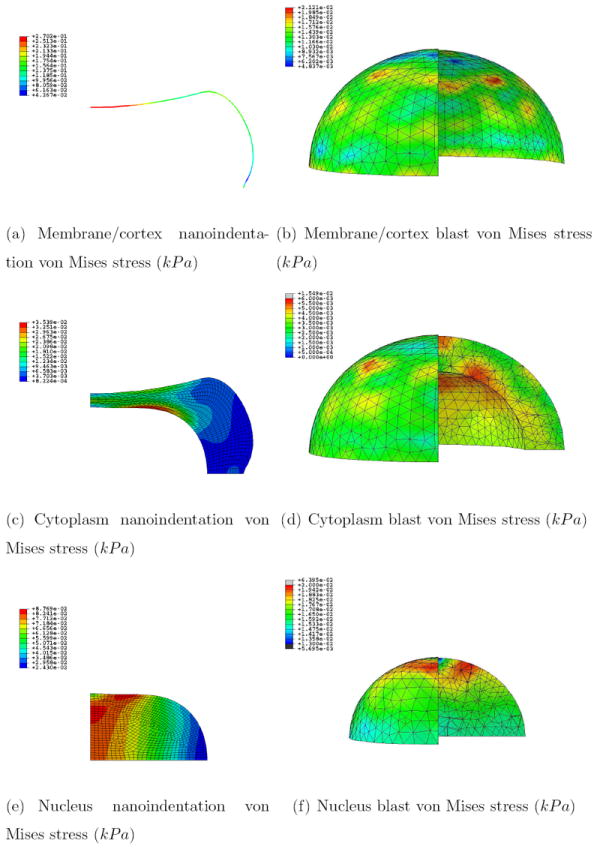
Traumatic brain injuries have recently been put under the spotlight as one of the most important causes of accidental brain dysfunctions. Significant experimental and modeling efforts are thus underway to study the associated biological, mechanical and physical mechanisms. In the field of cell mechanics, progress is also being made at the experimental and modeling levels to better characterize many of the cell functions, including differentiation, growth, migration and death. The work presented here aims to bridge both efforts by proposing a continuum model of a neuronal cell submitted to blast loading. In this approach, the cytoplasm, nucleus and membrane (plus cortex) are differentiated in a representative cell geometry, and different suitable material constitutive models are chosen for each one. The material parameters are calibrated against published experimental work on cell nanoindentation at multiple rates. The final cell model is ultimately subjected to blast loading within a complete computational framework of fluid-structure interaction. The results are compared to the nanoindentation simulation, and the specific effects of the blast wave on the pressure and shear levels at the interfaces are identified. As a conclusion, the presented model successfully captures some of the intrinsic intracellular phenomena occurring during the cellular deformation under blast loading that potentially lead to cell damage. It suggests, more particularly, that the localization of damage at the nucleus membrane is similar to what has already been observed at the overall cell membrane. This degree of damage is additionally predicted to be worsened by a longer blast positive phase duration. In conclusion, the proposed model ultimately provides a new three-dimensional computational tool to evaluate intracellular damage during blast loading.
Copyright © 2012 Acta Materialia Inc. All rights reserved.
Figures
Similar articles
-
Mechanics of blast loading on the head models in the study of traumatic brain injury using experimental and computational approaches.Biomech Model Mechanobiol. 2013 Jun;12(3):511-31. doi: 10.1007/s10237-012-0421-8. Epub 2012 Jul 26. Biomech Model Mechanobiol. 2013. PMID: 22832705
-
Investigation of wave propagation through head layers with focus on understanding blast wave transmission.Biomech Model Mechanobiol. 2020 Jun;19(3):875-892. doi: 10.1007/s10237-019-01256-9. Epub 2019 Nov 19. Biomech Model Mechanobiol. 2020. PMID: 31745681
-
Development of a finite element model for blast brain injury and the effects of CSF cavitation.Ann Biomed Eng. 2012 Jul;40(7):1530-44. doi: 10.1007/s10439-012-0519-2. Ann Biomed Eng. 2012. PMID: 22298329
-
Mechanisms of primary blast-induced traumatic brain injury: insights from shock-wave research.J Neurotrauma. 2011 Jun;28(6):1101-19. doi: 10.1089/neu.2010.1442. Epub 2011 May 5. J Neurotrauma. 2011. PMID: 21332411 Review.
-
Application of the Johnson-Cook plasticity model in the finite element simulations of the nanoindentation of the cortical bone.J Mech Behav Biomed Mater. 2020 Jan;101:103426. doi: 10.1016/j.jmbbm.2019.103426. Epub 2019 Sep 13. J Mech Behav Biomed Mater. 2020. PMID: 31557661 Review.
Cited by
-
Response of Single Cells to Shock Waves and Numerically Optimized Waveforms for Cancer Therapy.Biophys J. 2018 Mar 27;114(6):1433-1439. doi: 10.1016/j.bpj.2017.09.042. Biophys J. 2018. PMID: 29590600 Free PMC article.
-
Localized Axolemma Deformations Suggest Mechanoporation as Axonal Injury Trigger.Front Neurol. 2020 Jan 30;11:25. doi: 10.3389/fneur.2020.00025. eCollection 2020. Front Neurol. 2020. PMID: 32082244 Free PMC article.
-
A Multiscale Model to Predict Neuronal Cell Deformation with Varying Extracellular Matrix Stiffness and Topography.Cell Mol Bioeng. 2020 May 4;13(3):229-245. doi: 10.1007/s12195-020-00615-2. eCollection 2020 Jun. Cell Mol Bioeng. 2020. PMID: 32426060 Free PMC article.
-
The roles of cellular nanomechanics in cancer.Med Res Rev. 2015 Jan;35(1):198-223. doi: 10.1002/med.21329. Epub 2014 Aug 18. Med Res Rev. 2015. PMID: 25137233 Free PMC article. Review.
-
Visco-Hyperelastic Characterization of the Equine Immature Zona Pellucida.Materials (Basel). 2021 Mar 5;14(5):1223. doi: 10.3390/ma14051223. Materials (Basel). 2021. PMID: 33807624 Free PMC article.
References
-
- Khan M, Im Y-B, Shunmugavel A, Gilg AG, Dhindsa RK, Singh AK, Singh I. Administration of S-nitrosoglutathione after traumatic brain injury protects the neurovascular unit and reduces secondary injury in a rat model of controlled cortical impact. Journal of Neuroinflammation. 2009;6(32):1–12. doi: 10.1186/1742-2094-6-32. - DOI - PMC - PubMed
Publication types
MeSH terms
Grants and funding
LinkOut - more resources
Full Text Sources
Research Materials
