

Classical eyeblink conditioning using electrical stimulation of caudal mPFC as conditioned stimulus is dependent on cerebellar interpositus nucleus in guinea pigs
- PMID: 22562015
- PMCID: PMC4010383
- DOI: 10.1038/aps.2012.32
Classical eyeblink conditioning using electrical stimulation of caudal mPFC as conditioned stimulus is dependent on cerebellar interpositus nucleus in guinea pigs
Abstract
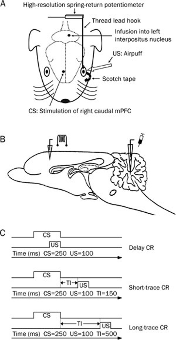
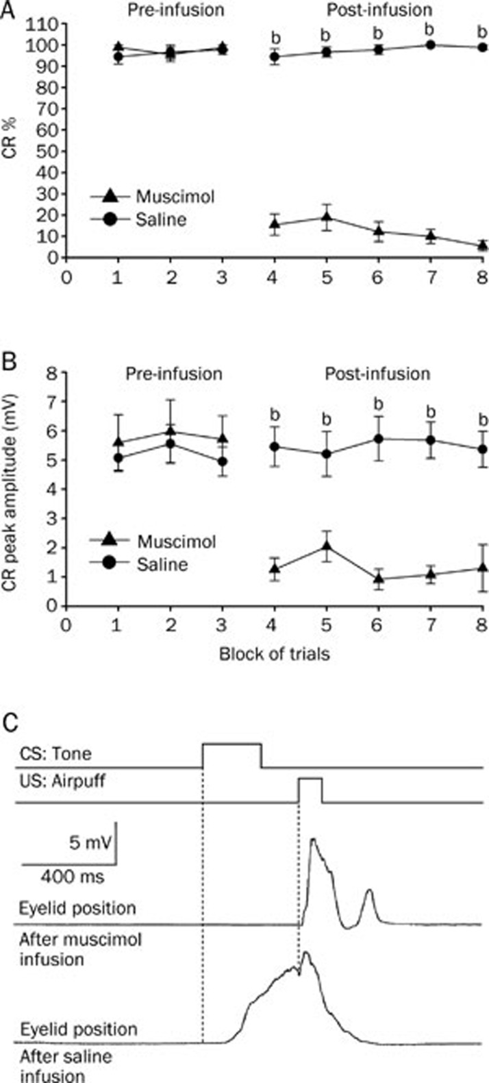
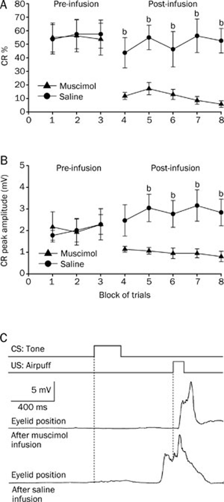
Aim: To determine whether electrical stimulation of caudal medial prefrontal cortex (mPFC) as conditioned stimulus (CS) paired with airpuff unconditioned stimulus (US) was sufficient for establishing eyeblink conditioning in guinea pigs, and whether it was dependent on cerebellar interpositus nucleus.
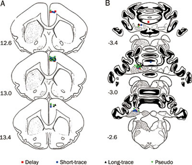
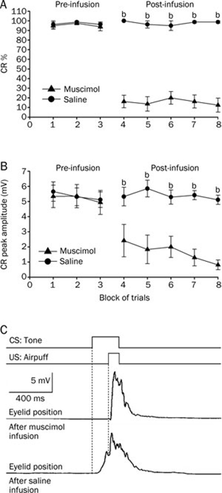
Methods: Thirty adult guinea pigs were divided into 3 conditioned groups, and trained on the delay eyeblink conditioning, short-trace eyeblink conditioning, and long-trace eyeblink conditioning paradigms, respectively, in which electrical stimulation of the right caudal mPFC was used as CS and paired with corneal airpuff US. A pseudo conditioned group of another 10 adult guinea pigs was given unpaired caudal mPFC electrical stimulation and the US. Muscimol (1 μg in 1 μL saline) and saline (1 μL) were infused into the cerebellar interpositus nucleus of the animals through the infusion cannula on d 11 and 12, respectively.
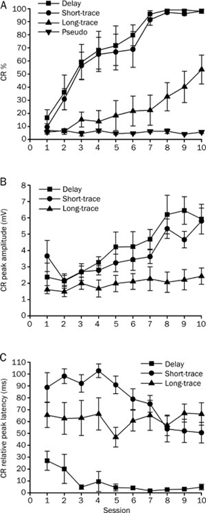
Results: The 3 eyeblink conditioning paradigms have been successfully established in guinea pigs. The animals acquired the delay and short-trace conditioned responses more rapidly than long-trace conditioned responses. Muscimol infusion into the cerebellar interpositus nucleus markedly impaired the expression of the 3 eyeblink conditioned responses.
Conclusion: Electrical stimulation of caudal mPFC is effective CS for establishing eyeblink conditioning in guinea pigs, and it is dependent on the cerebellar interpositus nucleus.
Figures
Similar articles
-
Disrupted topography of the acquired trace-conditioned eyeblink responses in guinea pigs after suppression of cerebellar cortical inhibition to the interpositus nucleus.Brain Res. 2010 Jun 14;1337:41-55. doi: 10.1016/j.brainres.2010.03.089. Epub 2010 Apr 8. Brain Res. 2010. PMID: 20381463
-
Transfer of classical eyeblink conditioning with electrical stimulation of mPFC or tone as conditioned stimulus in guinea pigs.Behav Brain Res. 2014 Nov 1;274:19-29. doi: 10.1016/j.bbr.2014.07.051. Epub 2014 Aug 10. Behav Brain Res. 2014. PMID: 25106738
-
Dissociaton of conditioned eye and limb responses in the cerebellar interpositus.Physiol Behav. 2007 May 16;91(1):9-14. doi: 10.1016/j.physbeh.2007.01.006. Epub 2007 Jan 20. Physiol Behav. 2007. PMID: 17320121
-
Role of the nuclei in eyeblink conditioning.Ann N Y Acad Sci. 2002 Dec;978:93-105. doi: 10.1111/j.1749-6632.2002.tb07558.x. Ann N Y Acad Sci. 2002. PMID: 12582044 Review.
-
The Anatomy and Physiology of Eyeblink Classical Conditioning.Curr Top Behav Neurosci. 2018;37:297-323. doi: 10.1007/7854_2016_455. Curr Top Behav Neurosci. 2018. PMID: 28025812 Review.
Cited by
-
Prefrontal control of cerebellum-dependent associative motor learning.Cerebellum. 2014 Feb;13(1):64-78. doi: 10.1007/s12311-013-0517-4. Cerebellum. 2014. PMID: 24013852
-
Electrical Stimulation Normalizes c-Fos Expression in the Deep Cerebellar Nuclei of Depressive-like Rats: Implication of Antidepressant Activity.Cerebellum. 2017 Apr;16(2):398-410. doi: 10.1007/s12311-016-0812-y. Cerebellum. 2017. PMID: 27435250
-
Trace eyeblink conditioning is associated with changes in synaptophysin immunoreactivity in the cerebellar interpositus nucleus in guinea pigs.Biosci Rep. 2018 May 8;38(3):BSR20170335. doi: 10.1042/BSR20170335. Print 2018 Jun 29. Biosci Rep. 2018. PMID: 29051391 Free PMC article.
-
Establishment and transfer of classical eyeblink conditioning using electrical microstimulation of the hippocampus as the conditioned stimulus.PLoS One. 2017 Jun 2;12(6):e0178502. doi: 10.1371/journal.pone.0178502. eCollection 2017. PLoS One. 2017. PMID: 28575003 Free PMC article.
-
Pretrial functional connectivity differentiates behavioral outcomes during trace eyeblink conditioning in the rabbit.Learn Mem. 2016 Mar 15;23(4):161-8. doi: 10.1101/lm.040220.115. Print 2016 Apr. Learn Mem. 2016. PMID: 26980784 Free PMC article.
References
-
- Christian KM, Thompson RF. Neural substrates of eyeblink conditioning: acquisition and retention. Learn Mem. 2003;10:427–55. - PubMed
-
- Christian KM, Thompson RF. Long-term storage of an associative memory trace in the cerebellum. Behav Neurosci. 2005;119:526–37. - PubMed
-
- Thompson RF. In search of memory traces. Annu Rev Psychol. 2005;56:1–23. - PubMed
Publication types
MeSH terms
Substances
LinkOut - more resources
Full Text Sources
