Review
. 2012 Aug;1821(8):1078-88.
doi: 10.1016/j.bbalip.2012.04.004. Epub 2012 Apr 22.

Regulation of the Golgi complex by phospholipid remodeling enzymes

Affiliations
Review

Regulation of the Golgi complex by phospholipid remodeling enzymes

Kevin D Ha et al. Biochim Biophys Acta. 2012 Aug.

Abstract

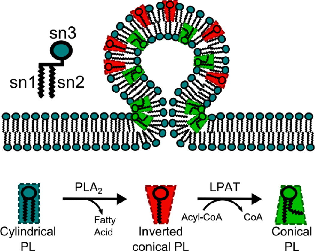
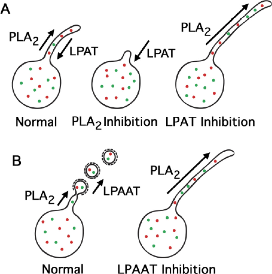
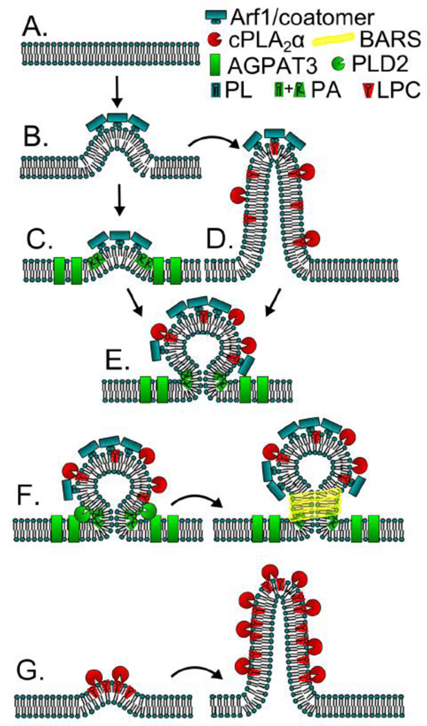
The mammalian Golgi complex is a highly dynamic organelle consisting of stacks of flattened cisternae with associated coated vesicles and membrane tubules that contribute to cargo import and export, intra-cisternal trafficking, and overall Golgi architecture. At the morphological level, all of these structures are continuously remodeled to carry out these trafficking functions. Recent advances have shown that continual phospholipid remodeling by phospholipase A (PLA) and lysophospholipid acyltransferase (LPAT) enzymes, which deacylate and reacylate Golgi phospholipids, respectively, contributes to this morphological remodeling. Here we review the identification and characterization of four cytoplasmic PLA enzymes and one integral membrane LPAT that participate in the dynamic functional organization of the Golgi complex, and how some of these enzymes are integrated to determine the relative abundance of COPI vesicle and membrane tubule formation. This article is part of a Special Issue entitled Lipids and Vesicular Transport.


Figures

Figure 1
Figure 1
A simple model of Lands cycle phospholipid remodeling by PLA2 and LPAT enzymes and how their phospholipid products can generate membrane curvature based on their physical shapes.
Figure 2
Figure 2
Membrane trafficking pathways from and within the mammalian Golgi complex. Several studies have recently reported that four cytoplasmic PLA enzymes are associated with the Golgi complex and regulate trafficking in various ways. As shown in Step 1, the cytoplasmic PLA2 enzyme PAFAHIb induces membrane tubules from the Golgi complex that contribute to the formation of an intact ribbon structure. In addition, PAFAHIb also appears to influence anterograde trafficking from the TGN (Step 2). A second cytoplasmic enzyme, cPLA2α, mediates the formation of intra-cisternal membrane tubules that appear to facilitate anterograde transport when the Golgi complex receives a large bolus of secretory cargo (Step 3). In addition, cPLA2 may also negatively regulate COPI coated vesicle formation (Fig. 2, Step 4), perhaps by shifting the balance toward membrane tubule formation. A third cytoplasmic PLA2, PLA2G6, was shown to mediate the formation of membrane tubules from the ERGIC, which may be important for connecting regions involved in COPI vesicle budding (Step 5). The final PLA enzyme recently found associated with ERGIC and Golgi membranes is iPLA1γ, which appears to contribute to anterograde transport through the Golgi complex by an unknown mechanism.
Figure 3
Figure 3
Possible routes for the individual and concerted actions of PLA2 and LPAT enzymes to influence the formation of membrane tubules and coated vesicles. (A) Membrane tubules could be generated and regulated by the opposing actions of PLA2 and LPAT enzymes. Hydrolysis of membrane phospholipids by cytoplasmic PLA2 enzymes could generate positive curvature inducing LPLs, ultimately resulting membrane tubule formation. The LPLs could be reacylated by LPAT enzymes back to phospholipids, thus negatively regulating the formation of membrane tubules. (B) The hydrolytic activity of cytoplasmic PLA2 enzymes could generate membrane tubules that are consumed through coated vesicle budding, which is facilitated by LPAAT activity for membrane fission. In this generic model, the coated vesicles could be either COPI vesicles budding from the ERGIC or AP-1 clathrin coated vesicles budding from the TGN. The consequences of inhibition by PLA2 and LPAT antagonists are shown, which have been supported by several studies (9, 66).
Figure 4
Figure 4
Models for cPLA2α and AGPAT3/LPAAT3 activities contributing to COPI vesicle and Golgi membrane tubule formation. (A) Flat membrane that will be modified by curve producing proteins; (B) Arf1 and coatomer binding deforms membranes, initiating the first steps of vesiculation or tubulation; (B to C) AGPAT3/LPAAT3 activity produces unsaturated PA, a lipid that resists positive curvature; (B to D) cPLA2α activity produces LPC, a positive curvature stabilizing and tubule inducing lipid; (C and D to E) LPC stabilizes positive curvature of the bud and unsaturated PA stabilizes negative curvature at the bud neck; (F) The concerted PA production activities of AGPAT3/LPAAT3 and PLD2 at the bud neck help to recruit BARS and aide vesicle fission; (G) PLA2 activity can induce membrane curvature and tubulation in the absence of Arf1 and coatomer.
Figure 5
Figure 5
Model integrating membrane curvature produced by the PLA2 activity of PAFAHIb α1 and α2 with Lis1-mediated dynein transport along microtubules. PAFAHIb initiates outward membrane curvature to generate a membrane tubule, which can be pulled/extended along microtubules (MT) by Lis1 and Ndel interactions with dynein. Dynein may be able to carry the membrane tubule towards the minus end of microtubules, facilitating the convergence of the Golgi stack and positioning at the microtubule organizing center (MTOC, minus end of the microtubules).
Figure 6
Figure 6
Enzymatic pathways, several involved in Lands cycle reactions, which lead to the modification and interconversion of lipid species. Lysophosphatidic acid (LPA); phosphatidic acid (PA); diacylglycerol (DAG); phosphatidylcholine (PC); lysophosphatidylcholine (LPC); sphingomyelin (SM); ceramide (Cer); phosphatidylinositol 4-phosphate (PI4P); phosphatidylinositol 4, 5-phosphate (PI4,5P); 1-acylglycerol-3-phosphate acyltransferase 3 (AGPAT3); lysophosphatidic acid acyltransferase 3 (LPAAT3); phospholipase A2 (PLA2); diacylglycerol kinase (DAGK); phosphatidic acid phosphatase (PA P’tase); phospholipase D (PLD); PC specific phospholipase C (PC-PLC); phosphatidylinositol specific PLC (PI-PLC); sphingomyelin synthase (SMS).

References

    1. Farquhar MG, Palade GE. The Golgi apparatus (complex)-(1954–1981)-from artifact to center stage. J Cell Biol. 1981;91:77s–103s. - PMC - PubMed
    1. Farquhar MG, Palade GE. The Golgi apparatus: 100 years of progress and controversy. Trends Cell Biol. 1998;8:2–10. - PMC - PubMed
    1. Lowe M. Structural organization of the Golgi apparatus. Curr Opin Cell Biol. 2011;23:85–93. - PubMed
    1. Polishchuk RS, Capestrano M, Polishchuk EV. Shaping tubular carriers for intracellular membrane transport. FEBS Lett. 2009;583:3847–3856. - PubMed
    1. Wilson C, Venditti R, Rega LR, Colanzi A, D'Angelo G, De Matteis MA. The Golgi apparatus: an organelle with multiple complex functions. Biochem J. 2011;433:1–9. - PubMed

Publication types

MeSH terms

LinkOut - more resources