. 2012 Jul 1;8(7):1032-44.
doi: 10.4161/auto.20123. Epub 2012 May 7.

Suppression of basal autophagy reduces lung cancer cell proliferation and enhances caspase-dependent and -independent apoptosis by stimulating ROS formation

Affiliations

Suppression of basal autophagy reduces lung cancer cell proliferation and enhances caspase-dependent and -independent apoptosis by stimulating ROS formation

Vitaliy O Kaminskyy et al. Autophagy. .

Abstract

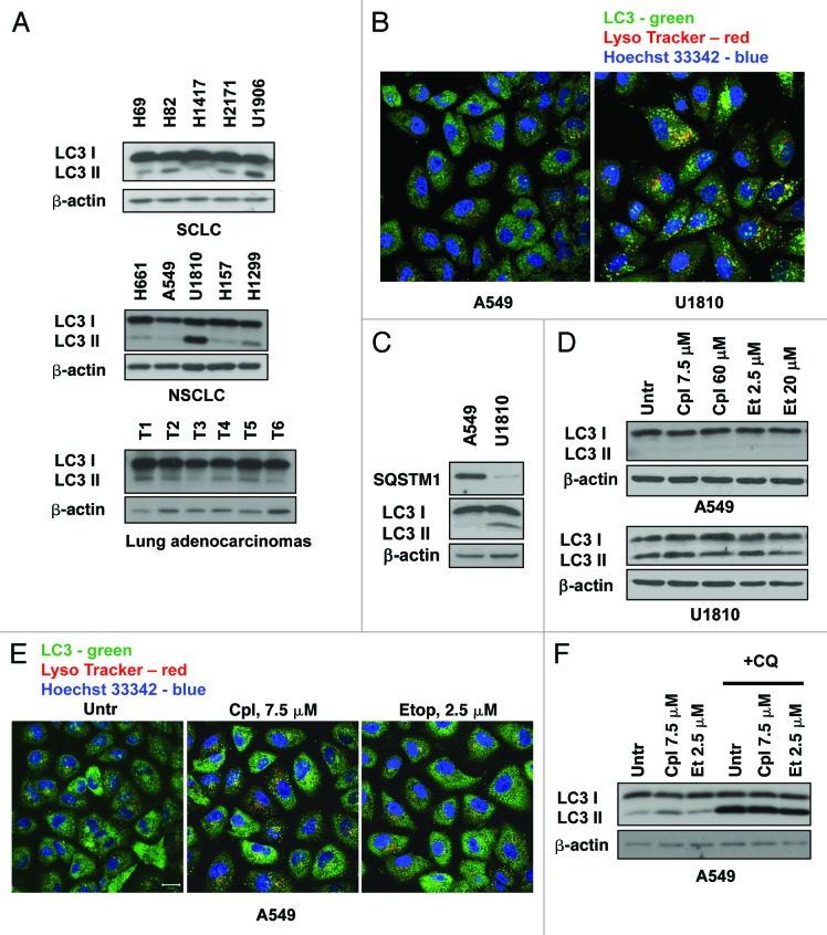
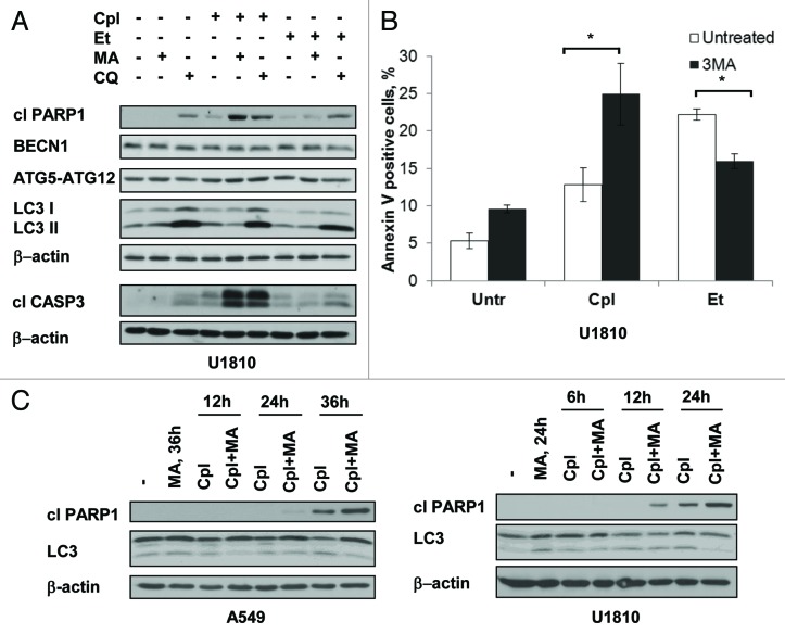
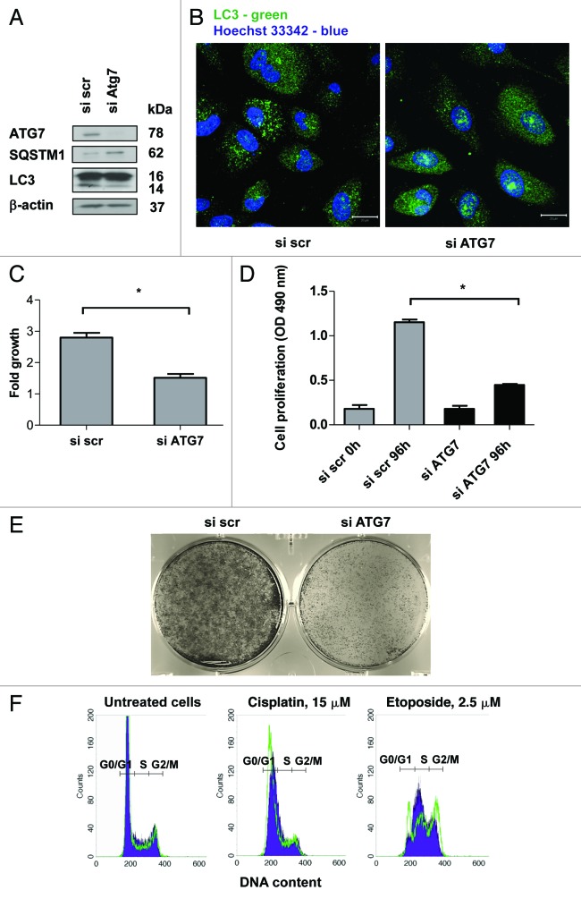
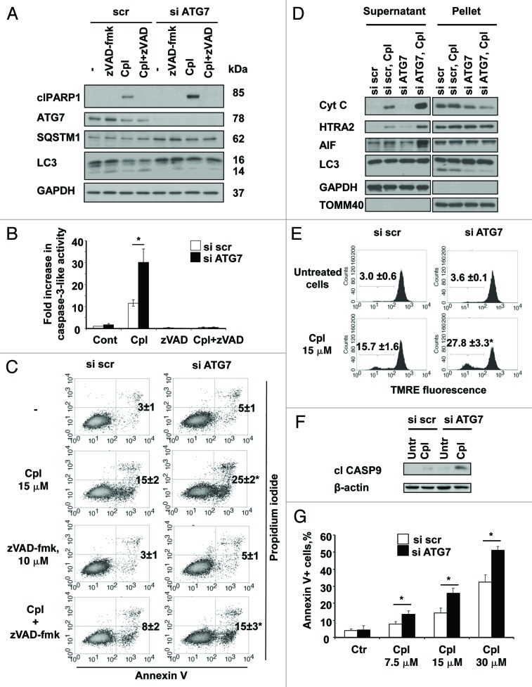
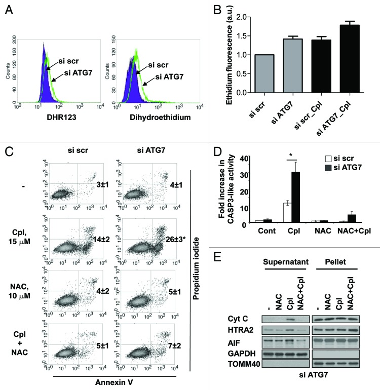
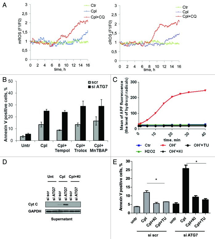
Autophagy is a catabolic process involved in the turnover of organelles and macromolecules which, depending on conditions, may lead to cell death or preserve cell survival. We found that some lung cancer cell lines and tumor samples are characterized by increased levels of lipidated LC3. Inhibition of autophagy sensitized non-small cell lung carcinoma (NSCLC) cells to cisplatin-induced apoptosis; however, such response was attenuated in cells treated with etoposide. Inhibition of autophagy stimulated ROS formation and treatment with cisplatin had a synergistic effect on ROS accumulation. Using genetically encoded hydrogen peroxide probes directed to intracellular compartments we found that autophagy inhibition facilitated formation of hydrogen peroxide in the cytosol and mitochondria of cisplatin-treated cells. The enhancement of cell death under conditions of inhibited autophagy was partially dependent on caspases, however, antioxidant NAC or hydroxyl radical scavengers, but not the scavengers of superoxide or a MnSOD mimetic, reduced the release of cytochrome c and abolished the sensitization of the cells to cisplatin-induced apoptosis. Such inhibition of ROS prevented the processing and release of AIF (apoptosis-inducing factor) and HTRA2 from mitochondria. Furthermore, suppression of autophagy in NSCLC cells with active basal autophagy reduced their proliferation without significant effect on the cell-cycle distribution. Inhibition of cell proliferation delayed accumulation of cells in the S phase upon treatment with etoposide that could attenuate the execution stage of etoposide-induced apoptosis. These findings suggest that autophagy suppression leads to inhibition of NSCLC cell proliferation and sensitizes them to cisplatin-induced caspase-dependent and -independent apoptosis by stimulation of ROS formation.

Keywords: NSCLC; ROS; apoptosis; autophagy; caspase-independent cell death; hydroxyl radical; superoxide.


Figures

None
Figure 1. Autophagy in lung cancer cell lines and its activity upon treatment with cisplatin and etoposide. (A) Different level of LC3 lipidation in lung carcinoma cell lines and human lung adenocarcinomas. (B) Co-staining with LC3 antibody and Lysotracker Red shows significant numbers of autophagosomes in U1810 cells fused with lysosomes. (C) Expression of SQSTM1 protein and lipidation of LC3 in A549 and U1810 cells detected by immunoblotting. (D) Expression of LC3 in U1810 and A549 cells treated (24 h) with cisplatin (7.5 or 60 μM) or etoposide (2.5 and 20 μM). (E) Immunocytochemical detection of autophagosome formation in A549 cells treated (24 h) with cisplatin (7.5 μM) or etoposide (2.5 μM). (F) Autophagic flux in A549 cells treated (36 h) with etoposide (2.5 μM) or cisplatin (7.5 μM). Cells were treated with chloroquine (CQ, 50 μM) 3 h before collecting the samples and accumulation of LC3-II was measured by immunoblotting.
None
Figure 2. The inhibitors of autophagy sensitize NSCLC cells to cisplatin-induced apoptosis and attenuate the cell death induced by etoposide. (A) Activation of CASP3, cleavage of PARP1 and expression of BECN1, ATG7, ATG5–12 and LC3 in U1810 cells treated (24 h) with cisplatin (Cpl, 15 μM) or etoposide (Et, 2.5 μM) alone or in combination with either inhibitors of autophagy 3-methyladenine (3MA, 5 mM) or chloroquine (CQ, 50 μM). (B) Annexin V/ PI staining of U1810 cells treated (24 h) with cisplatin (15 μM) or etoposide (2.5 μM) alone, or in combination with 3-methyladenine. (C) Time-course analysis of PARP1 cleavage, expression of LC3 in A549 and U1810 cells treated with cisplatin (15 μM), 3-methyladenine (5 mM), or their combination. Statistical significance: *p < 0.05
None
Figure 3. The inhibition of autophagy delays the growth and progression of cells through the cell cycle. (A) Inhibition of autophagy in U1810 cells transfected with siRNA targeting ATG7. Autophagy was detected by measurement of the expression level of SQSTM1 and conversion of LC3-I to LC3-II proteins, or (B) using immunostaining of autophagosomes with LC3 antibody. (C) Autophagy inhibition delays the growth of U1810 cells. The cells were incubated (24 h) with scrambled or ATG7-targeting siRNAs. The cells were plated in 6-well plates and counted 48h after seeding. The bars represent a fold increase in the number of seeded cells. (D) Suppression of autophagy inhibits cell proliferation. U1810 cells were plated in 96-well plates and MTS assay was performed 96h after seeding. (E) Suppression of autophagy inhibits colony formation in U1810 cells. Autophagy was suppressed using siRNA targeting ATG7. The cells were grown in 6-well plates and five days after fixed and stained with crystal violet. (F) The cell cycle analysis of U1810 cells with basal or suppressed autophagy was performed after treatment with cisplatin (15 μM), or etoposide (2.5 μM). The filled blue histogram represents the cells transfected with scrambled siRNA and the green histogram corresponds to the cells with suppressed autophagy. Statistical significance: *p < 0.05
None
Figure 4. The inhibition of autophagy sensitizes the cells to caspase-dependent and -independent apoptotic cell death induced by cisplatin. (A) U1810 cells with basal or siRNA-mediated suppressed autophagy were pretreated (1 h) with DMSO or pan-caspase inhibitor zVAD-fmk (10 μM) and were then treated with cisplatin (15 μM). The level of autophagy was measured by the detection of expression of SQSTM1 and LC3 proteins by western blot and the efficiency of effector caspases inhibition was proved by the detection of PARP1 cleavage and (B) a caspase activity assay. (C) U1810 cells were transfected with scrambled or siRNA-targeting ATG7, and were then pretreated (1 h) with zVAD-fmk (10 μM) followed by cisplatin administration (15 μM, 24 h). Cell death was measured by annexin V/PI-staining. The number of apoptotic/secondary necrotic cells is shown as the sum of annexin V-positive and annexin V/PI double-positive cells. (D) The effect of autophagy inhibition on the distribution of cytochrome c, HTRA2 and AIF in the cytosolic and membrane fractions of U1810 cells treated with cisplatin. (E) The effect of autophagy inhibition on mitochondrial membrane potential in U1810 cells treated with cisplatin. The cells were treated (24 h) with cisplatin and MMP was assessed by TMRE staining. (F) Activation of CASP9 in cells with basal and suppressed autophagy. U1810 cells were treated (24 h) with cisplatin and the active form of CASP9 was detected by immunoblotting. (G) The effect of autophagy inhibition on cell death induced by different concentrations of cisplatin (7.5–30 μM). U1810 cells were treated (24 h) with cisplatin and cell death was measured by Annexin V/PI-staining. Statistical significance: *p < 0.05–untreated or treated cells transfected with scrambled siRNA in comparison to the untreated control (or same cell treatment) transfected with siRNA targeting ATG7.
None
Figure 5. The inhibition of autophagy stimulates ROS formation which is required for sensitization of NSCLC cells to cisplatin-induced apoptosis. (A) ROS formation in U1810 cells transfected with scrambled or ATG7-targeting siRNA. Forty-eight hours after transfection ROS formation was measured using dihydrorodamine 123 and dihydroethidium probes, as described in the Materials and Methods. (B) The effect of autophagy inhibition on ROS formation in U1810 cells. Cells were transfected with scrambled or ATG7-targeting siRNAs followed by treatment with cisplatin (15 μM, 24 h). (C) U1810 cells with basal or suppressed autophagy were pretreated (1 h) with N-acetyl-cysteine (10 mM) and then incubated with cisplatin (15 μM, 24 h). Cell death was measured by Annexin V/PI staining and (D) the CASP3-like activity assay was performed as described in the Materials and Methods. (E) The effect of the antioxidant NAC on cisplatin-mediated release of cytochrome c and AIF from mitochondria in U1810 cells with suppressed autophagy. Cells were treated as described in (C). Statistical significance: *p < 0.05.
None
Figure 6. The scavengers of hydroxyl radicals prevent cisplatin-mediated cell death in NSCLC cells with suppressed autophagy. (A) Detection of H2O2 formation in cytosolic and mitochondrial compartments of U1810 cells treated with cisplatin (15 μM) alone or in combination with chloroquine (50 μM). Representative single cells that responded to treatment from different live cell imaging experiments are shown. (B) The effect of the MnSOD mimetic MnTBAP and scavengers of superoxide Tempol and Trolox on cisplatin-mediated cell death in U1810 cells with suppressed autophagy. Autophagy was suppressed in U1810 cells using siRNA targeting ATG7 and 48 h after transfection cells were pretreated for 1 h with Trolox (200 μM), Tempol (0.5 mM) or MnTBAP (25 μM) followed by treatment with cipslatin (15 μM, 24 h). (C) Thiourea and potassium iodide efficiently scavenge hydroxyl radicals in vitro. (D) The scavengers of hydroxyl radical potassium iodide (5 mM) and thiourea (1 mM) prevent the release of cytochrome c and processing of AIF in U1810 cells treated with cisplatin (15 μM, 24 h). Cells were pretreated for 1 h with the scavengers of hydroxyl radicals followed by treatment with cisplatin. (E) The effect of hydroxyl radical scavengers on cell death induced by cisplatin in U1810 cells with basal or suppressed autophagy. Cells were treated as described in (D). Statistical significance: *p < 0.05.

Similar articles

Cited by

References

    1. Narendra D, Tanaka A, Suen DF, Youle RJ. Parkin is recruited selectively to impaired mitochondria and promotes their autophagy. J Cell Biol. 2008;183:795–803. doi: 10.1083/jcb.200809125. - DOI - PMC - PubMed
    1. Yousefi S, Perozzo R, Schmid I, Ziemiecki A, Schaffner T, Scapozza L, et al. Calpain-mediated cleavage of Atg5 switches autophagy to apoptosis. Nat Cell Biol. 2006;8:1124–32. doi: 10.1038/ncb1482. - DOI - PubMed
    1. Wirawan E, Vande Walle L, Kersse K, Cornelis S, Claerhout S, Vanoverberghe I, et al. Caspase-mediated cleavage of Beclin-1 inactivates Beclin-1-induced autophagy and enhances apoptosis by promoting the release of proapoptotic factors from mitochondria. Cell Death Dis 2010; 1. - PMC - PubMed
    1. Eisenberg-Lerner A, Bialik S, Simon HU, Kimchi A. Life and death partners: apoptosis, autophagy and the cross-talk between them. Cell Death Differ. 2009;16:966–75. doi: 10.1038/cdd.2009.33. - DOI - PubMed
    1. Pyo JO, Nah J, Kim HJ, Lee HJ, Heo J, Lee H, et al. Compensatory activation of ERK1/2 in Atg5-deficient mouse embryo fibroblasts suppresses oxidative stress-induced cell death. Autophagy. 2008;4:315–21. - PubMed

Publication types

MeSH terms