. 2012 Nov;61(11):2079-90.
doi: 10.1007/s00262-012-1272-y. Epub 2012 May 6.

Uptake routes of tumor-antigen MAGE-A3 by dendritic cells determine priming of naïve T-cell subtypes

Affiliations

Uptake routes of tumor-antigen MAGE-A3 by dendritic cells determine priming of naïve T-cell subtypes

Ines Moeller et al. Cancer Immunol Immunother. 2012 Nov.

Abstract

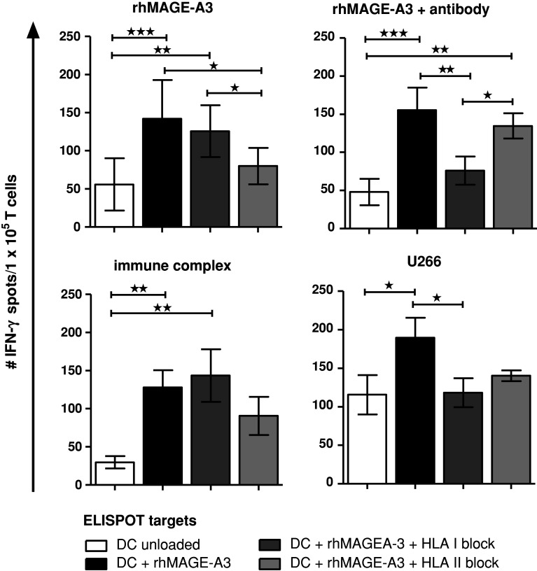
Induction of tumor-antigen-specific T cells in active cancer immunotherapy is generally difficult due to the very low anti-tumoral precursor cytotoxic T cells. By improving tumor-antigen uptake and presentation by dendritic cells (DCs), this problem can be overcome. Focusing on MAGE-A3 protein, frequently expressed in many types of tumors, we analyzed different DC-uptake routes after additional coating the recombinant MAGE-A3 protein with either a specific monoclonal antibody or an immune complex formulation. Opsonization of the protein with antibody resulted in increased DC-uptake compared to the uncoated rhMAGE-A3 protein. This was partly due to Fcγ receptor-dependent internalization. However, unspecific antigen internalization via macropinocytosis also played a role. When analyzing DC-uptake of MAGE-A3 antigen expressed in multiple myeloma cell line U266, pretreatment with proteasome inhibitor bortezomib resulted in increased apoptosis compared to γ-irradiation. Bortezomib-mediated immunogenic apoptosis, characterized by elevated surface expression of hsp90, triggered higher phagocytosis of U266 cells by DCs involving specific DC-derived receptors. We further investigated the impact of antigen delivery on T-cell priming. Induction of CD8(+) T-cell response was favored by stimulating naïve T cells with either antibody-opsonized MAGE-A3 protein or with the bortezomib-pretreated U266 cells, indicating that receptor-mediated uptake favors cross-presentation of antigens. In contrast, CD4(+) T cells were preferentially induced after stimulation with the uncoated protein or protein in the immune complex, both antigen formulations were preferentially internalized by DCs via macropinocytosis. In summary, receptor-mediated DC-uptake mechanisms favored the induction of CD8(+) T cells, relevant for clinical anti-tumor response.


Conflict of interest statement

The authors declare no potential conflict of interest.

Figures

Fig. 1
Fig. 1
a Clinical course of a HLA-A1-positive and MAGE-A3 expressing malignant melanoma patient. ED first diagnosis, DLI donor lymphocyte infusion, formula image 
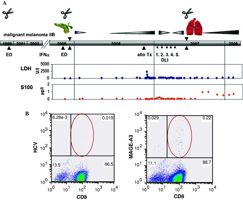
surgery. b Blood was collected after allogeneic PBSCT and stimulated twice with peptide EVDPIGHLY (MAGE-A3/HLA-A1). Mage-A3-specific T-cells were detected by pentamer assay. HCV peptide as negative control
Fig. 2
Fig. 2
Bortezomib significantly enhances apoptosis and U266 phagocytosis by DCs compared to γ-irradiation: a U266 cells, pretreated with either 30 Gy or 100 nM bortezomib (24 h), were stained with AnnexinV; n = 5. b hsp90 surface expression of apoptotic U266 cells pretreated as indicated. hsp90 inhibitor geldanamycin was applied 24 h before γ-irradiation or concurrently to bortezomib culture; n = 3. c Immature DCs (CD11c+) were loaded with U266 cells prepared in (b) (stained with CellTracker™) and co-cultured for 24 h at 37 °C. As indicated DC-derived CD91 were blocked by comparative inhibition adding 100 μM BSA and α2-macroglobulin, respectively. CD11c+/green double-positive cells are shown; n = 3. Cells were measured by flow cytometry. Data represent mean ± SEM of independent experiments; (a + b) one-way ANOVA with Bonferroni’s multiple comparison test; c Friedman test, with Dunn’s multiple comparison test; *p < 0.05, **p < 0.01, ***p < 0.001
Fig. 3
Fig. 3
Different affinities of anti-MAGE antibodies to FcγR: a Influence of IgG isotype on receptor affinity: Binding of murine antibody-opsonized MAGE-A3 protein to different Fcγ receptors (FcγR) expressed on THP-1 cell-line and primary NK cells, respectively, measured by flow cytometry. Negative control was loaded MAGE-A3 protein. Antibody concentrations: 0.2 μg/ml (dotted), 2 μg/ml (dashed), 20 μg/ml (solid). b Antigen binding was shown by intracellular staining of serial antibody dilutions of either normal (gly) or deglycosylated (de-gly) antibodies in MAGE-A3 expressing U266 cells, measured by flow cytometry. MFI values of one representative out of 3 independent experiments. Curves describe non-linear one site specific binding. c Anti-MAGE antibodies 6C1 and 57B were deglycosylated using PNGase F, cleaving Fc residue Asn297. Original antibody (gly); deglycosylated antibody (de-gly). One representative out of three experiments is shown. Antibody concentrations: 0.2 μg/ml (dotted), 2 μg/ml (dashed), 20 μg/ml (solid)
Fig. 4
Fig. 4
Opsonization of rhMAGE-A3 results in increased DC-uptake levels, significantly mediated by FcγR. a Expression of FcγR of immature DCs. Bars indicate mean ± SEM of n = 62 independent experiments of 54 different donors. Significant differences within indicated receptors were calculated using Friedman test with Dunn’s multiple comparison test, *p < 0.05, **p < 0.01, ***p < 0.001. b Immature DCs (CD11c+) were loaded in parallel with three different rhMAGE-A3-FITC antigen formulation for 30 min. CD11c+/FITC double-positive cells were measured by digital imaging system Scan^R and blotted on the y-axis; FcγR uptake was neutralized by specific antibody, macropinocytosis was inhibited by rottlerin. Data represent relative DC-uptake compared to the unloaded rhMAGE-A3 protein uptake (100 %) of 7 independent experiments; mean ± SEM; paired t-test. Left rhMAGE-A3 protein; middle rhMAGE-A3 opsonized with anti-MAGE monoclonal antibody 6C1; right rhMAGE-A3 opsonized with anti-MAGE antibody and cross-linked with anti-light chain F(ab)2 (IC immune complex). c Immature DCs (CD11c+) were loaded with rhMAGE-A3 opsonized with either unmodified or deglycosylated 6C1 IgG2a antibody. Data represent relative DC-uptake compared to unloaded rhMAGE-A3 protein uptake (100 %) of 5 independent experiments; mean ± SEM; paired t-test
Fig. 5
Fig. 5
Naive T-cell priming against MAGE-A3 antigens depends on the mode of antigen delivery. Naive T cells were stimulated with immature DCs loaded with rhMAGE-A3 protein, opsonized rhMAGE-A3 antibody (6C1 IgG2a), rhMAGE-A3-immune complex and MAGE-A3-expressing U266 cells, pretreated with bortezomib. After two stimulation rounds INF-γ secretion of primed T cells was detected by ELISPOT. As ELISPOT targets, DCs were loaded with rhMAGE-A3 protD-fusion-protein, control rhMAGE-A3 protein and a MAGE-A3 peptide mix containing immunogenic HLA class I and class II epitopes, respectively. IFN-γ response of T cells is calculated by the mean of the DC/T-cells-derived spots subtracted by background spots. Shown are mean ± SEM of 4 independent experiments, 1-way ANOVA with Bonferroni’s multiple comparison test, *p < 0.05, **p < 0.01, ***p < 0.001
Fig. 6
Fig. 6
Discrimination between CD4+ and CD8+ T-cell IFN-γ secretion. Before loading DCs with the different ELISPOT targets, HLA-molecules were blocked using neutralizing antibodies against HLA class I and class II, respectively. T-cell response of one donor is demonstrated. Bars represent mean ± SD, values of statistically significant differences between response of T cells to rhMAGE-A3 antigen and corresponding blocked antigen was analyzed by 1-way ANOVA with Bonferroni’s multiple comparison test, *p < 0.05, **p < 0.01, ***p < 0.001; n = 1. Similar results have been obtained with different donors. T cells initially primed with the antibody-opsonized rhMAGE-A3 protein or the bortezomib-pretreated U266 cell line preferentially induced a CD8+ T-cell response, as demonstrated by decreased IFN-γ secretion after blocking with HLA class I antibodies, whereas T cells initially primed with the unloaded protein or the immune complex formulation favored a CD4+ T-cell response, demonstrated by reduced IFN-γ secretion after blocking the antigen presenting cells with HLA class II antibodies

References

    1. Van den Eynde BJ, van der Bruggen P. T cell defined tumor antigens. Curr Opin Immunol. 1997;9:684–693. doi: 10.1016/S0952-7915(97)80050-7. - DOI - PubMed
    1. Atanackovic D, Luetkens T, Hildebrandt Y, Arfsten J, Bartels K, et al. Longitudinal analysis and prognostic effect of cancer-testis antigen expression in multiple myeloma. Clin Cancer Res. 2009;15:1343–1352. doi: 10.1158/1078-0432.CCR-08-0989. - DOI - PubMed
    1. Yang B, O’Herrin SM, Wu J, Reagan-Shaw S, Ma Y, et al. MAGE-A, mMage-b, and MAGE-C proteins form complexes with KAP1 and suppress p53-dependent apoptosis in MAGE-positive cell lines. Cancer Res. 2007;67:9954–9962. doi: 10.1158/0008-5472.CAN-07-1478. - DOI - PubMed
    1. Laduron S, Deplus R, Zhou S, Kholmanskikh O, Godelaine D, et al. MAGE-A1 interacts with adaptor SKIP and the deacetylase HDAC1 to repress transcription. Nucleic Acids Res. 2004;32:4340–4350. doi: 10.1093/nar/gkh735. - DOI - PMC - PubMed
    1. Monte M, Simonatto M, Peche LY, Bublik DR, Gobessi S, et al. MAGE-A tumor antigens target p53 transactivation function through histone deacetylase recruitment and confer resistance to chemotherapeutic agents. Proc Natl Acad Sci USA. 2006;103:11160–11165. doi: 10.1073/pnas.0510834103. - DOI - PMC - PubMed

Publication types

MeSH terms