. 2012 Jun 8;53(7):3431-40.
doi: 10.1167/iovs.12-9432.

Antidiabetic drug metformin suppresses endotoxin-induced uveitis in rats

Affiliations

Antidiabetic drug metformin suppresses endotoxin-induced uveitis in rats

Nilesh M Kalariya et al. Invest Ophthalmol Vis Sci. .

Abstract

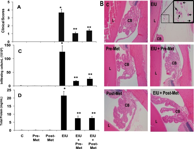
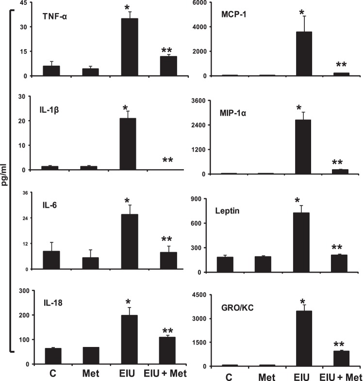
Purpose: To investigate the therapeutic effects of metformin, a commonly used antidiabetic drug, in preventing endotoxin-induced uveitis (EIU) in rats.

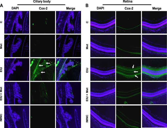
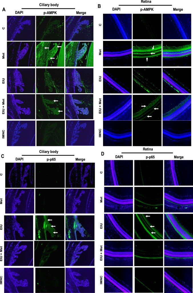
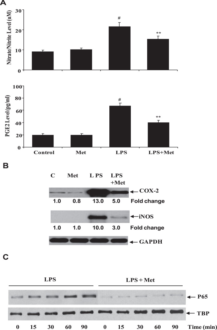
Methods: EIU in Lewis rats was developed by subcutaneous injection of lipopolysaccharide (LPS; 150 μg). Metformin (300 mg/kg body weight, intraperitoneally) or its carrier was injected either 12 hours before or 2 hours after LPS induction. Three and 24 hours after EIU, eyes were enucleated and aqueous humor (AqH) was collected. The MILLIPLEX-MAG Rat cytokine-chemokine magnetic bead array was used to determine inflammatory cytokines. The expression of Cox-2, phosphorylation of AMPK, and NF-κB (p65) were determined immunohistochemically. Primary human nonpigmented ciliary epithelial cells (HNPECs) were used to determine the in vitro efficacy of metformin.

Results: Compared with controls, the EIU rat AqH had significantly increased number of infiltrating cells and increased levels of various cytokines and chemokines (TNF-α, MCP-1, IL-1β, MIP-1α, IL-6, Leptin, and IL-18) and metformin significantly prevented the increase. Metformin also prevented the expression of Cox-2 and phosphorylation of p65, and increased the activation of AMPK in the ciliary bodies and retinal tissues. Moreover, metformin prevented the expression of Cox-2, iNOS, and activation of NF-kB in the HNPECs and decreased the levels of NO and PGE2 in cell culture media.

Conclusions: Our results for the first time demonstrate a novel role of the antidiabetic drug, metformin, in suppressing uveitis in rats and suggest that this drug could be developed to prevent uveitis complications.


Conflict of interest statement

Disclosure: N.M. Kalariya, None; M. Shoeb, None; N.H. Ansari, None; S.K. Srivastava, None; K.V. Ramana, None

Figures

Figure 1.
Figure 1.
Chemical structure of metformin.
Figure 2.
Figure 2.
Metformin prevents LPS-induced infiltration of inflammatory cells and increase in protein levels in AqH. (A) The pathologic score of EIU in Lewis rat eyes injected with LPS in the absence and presence of metformin was determined at 24 hours with a slit lamp microscope. Results are given as mean ± SD (n = 6). #P < 0.001 versus control. **P < 0.001 versus EIU (Wilcoxon–Mann-Whitney test). (B) Histopathological results of paraffin-embedded sections showing infiltrated cells (inset) in the anterior chamber of EIU rat eyes without or with metformin injected 12 hours before or 2 hours after LPS administration. H&E-stained serial sections of rat eyes were photographed under a light microscope. Magnification, ×200. (C) The infiltrated inflammatory cells were determined by trypan blue exclusion cell counting and (D) total protein levels in the AqH. Results are expressed as the mean ± SD (n = 5); *P < 0.001 versus the control group; **P < 0.05 versus the EIU group. C, control; Pre-Met, pretreatment with metformin; Post-Met, posttreatment with metformin; EIU, endotoxin-induced uveitis; EIU + Pre-Met, endotoxin-induced uveitis + pretreatment with metformin; EIU + Post-Met, endotoxin-induced uveitis + post treatment with metformin; CB, ciliary body; L, lens.
Figure 3.
Figure 3.
Posttreatment with metformin suppressed LPS-induced increase in cytokines and chemokines in AqH. The MILLIPLEX MAG Rat cytokine/chemokine magnetic bead panel along with Luminex xMAP detection method was used to determine cytokines and chemokines. Results are expressed as the mean ± SD (n = 3; AqH was pooled from two rats for each data point); *P < 0.01 versus the control group; **P < 0.05 versus the EIU group. The results are expressed as pg/mL. C, control; Met, metformin; EIU, endotoxin-induced uveitis; EIU + Met, endotoxin-induced uveitis + metformin.
Figure 4.
Figure 4.
Posttreatment with metformin prevents expression of Cox-2. Serial sections of paraformaldehyde-fixed rat eyes enucleated 24 hours after EIU induction were immunostained with antibodies against Cox-2 and pictures were taken using a Nikon epifluorescence microscope. Arrows indicate Cox-2 expression in (A) ciliary bodies as well as (B) retinal tissues. A representative image is shown (n = 4). C, control; Met, metformin; EIU, endotoxin-induced uveitis; EIU + Met, endotoxin-induced uveitis + metformin; IMNC, isotype-matched negative control. Magnification ×200.
Figure 5.
Figure 5.
Posttreatment with metformin triggers phosphorylation of AMPK (A, B) and suppresses p-65 phosphorylation (C, D) in ocular tissues of LPS-induced EIU. Serial sections of paraformaldehyde-fixed rat eyes enucleated 3 hours after EIU induction were immunostained with antibodies against phospho-AMPK (A, B) and phospho-p65 (C, D). Nuclei were stained with DAPI and pictures were taken by Nikon epifluorescence microscope. Representative images are shown (n = 4). Arrows indicate phosphorylated AMPK (A, B) and phospho-p65 (C, D) in ciliary body (A, C), as well as retinal tissues (B, D). C, control; Met, metformin; EIU, endotoxin-induced uveitis; EIU + Met, endotoxin-induced uveitis + metformin; IMNC, isotype-matched negative control. Magnification ×200.
Figure 6.
Figure 6.
Metformin prevents inflammatory response in HNPECs stimulated with LPS. (A) The levels of nitrate/nitrite and PGE2 in the culture media were determined with ELISA kit. Data are expressed as mean ± SD (n = 6). *P < 0.001 versus the control group; **P < 0.05 versus the LPS group. (B) The expression of Cox-2 and iNOS from cell lysates was determined by Western blot using specific antibodies. (C) The nuclear translocation of p65 was determined in the nuclear extract by Western blot using specific antibodies against p65. TBP, TATA binding protein (loading control); C, control; Met, metformin.

References

    1. Nussenblatt RB. The natural history of uveitis. Int Ophthalmol. 1990;14:303–308 - PubMed
    1. Rosenbaum JT, McDevitt HO, Guss RB, Egbert PR. Endotoxin-induced uveitis in rats as a model for human disease. Nature. 1980;286:611–613 - PubMed
    1. Sijssens KM, Rijkers GT, Rothova A, et al. Distinct cytokine patterns in the aqueous humor of children, adolescents and adults with uveitis. Ocul Immunol Inflamm. 2008;16:211–216 - PubMed
    1. Curnow SJ, Murray PI. Inflammatory mediators of uveitis: cytokines and chemokines. Curr Opin Ophthalmol. 2006;17:532–537 - PubMed
    1. Yadav UC, Srivastava SK, Ramana KV. Aldose reductase inhibition prevents endotoxin-induced uveitis in rats. Invest Ophthalmol Vis Sci. 2007;48:4634–4642 - PMC - PubMed

Publication types

MeSH terms