Comparative Study
. 2012 Aug 1;95(3):300-7.
doi: 10.1093/cvr/cvs160. Epub 2012 May 4.

Increased late sodium current contributes to long QT-related arrhythmia susceptibility in female mice

Affiliations
Comparative Study

Increased late sodium current contributes to long QT-related arrhythmia susceptibility in female mice

John S Lowe et al. Cardiovasc Res. .

Abstract

Aims: Female gender is a risk factor for long QT-related arrhythmias, but the underlying mechanisms remain uncertain. Here, we tested the hypothesis that gender-dependent function of the post-depolarization 'late' sodium current (I(Na-L)) contributes.

Methods and results: Studies were conducted in mice in which the canonical cardiac sodium channel Scn5a locus was disrupted, and expression of human wild-type SCN5A cDNA substituted. Baseline QT intervals were similar in male and female mice, but exposure to the sodium channel opener anemone toxin ATX-II elicited polymorphic ventricular tachycardia in 0/9 males vs. 6/9 females. Ventricular I(Na-L) and action potential durations were increased in myocytes isolated from female mice compared with those from males before and especially after treatment with ATX-II. Further, ATX-II elicited potentially arrhythmogenic early afterdepolarizations in myocytes from 0/5 male mice and 3/5 female mice.

Conclusion: These data identify variable late I(Na) as a modulator of gender-dependent arrhythmia susceptibility.


Figures

Figure 1
Figure 1
ECG tracings prior to and following ATX-II injection in one female mouse. (A) Baseline recording showing normal sinus rhythm and no discernable QT interval. (B) Following ATX-II injection, there is marked deformity of the T wave. (C) This is followed by development of polymorphic ventricular tachycardia.
Figure 2
Figure 2
QT measurement in male and female mice after ATX-II injection. (A) T wave amplitude measurement is marked with a double-headed arrow. (B) Quantification of the T wave is performed by marking the T90 area and counting pixels within the region. P < 0.05, *P < 0.01. ATX-II treatment produced much greater changes in both indices of repolarization in female compared with male animals.
Figure 3
Figure 3
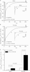
Late sodium current (INa-L) in male and female ventricular myocytes. Late sodium current in male (A) or female (B) myocytes with or without the addition of ATX-II. Female myocytes have a significantly larger late current at baseline that is further increased by ATX-II treatment. Late current was measured as a percentage of peak current before the ending of 200 ms pulsing after peak INa (indicated by arrows), while non-normalized raw current traces are shown in insets. Late current data (n = 6 each) are summarized in (C).
Figure 4
Figure 4
Comparisons of ventricular action potentials (APs) in male and female mice with and without ATX-II at a stimulation frequency of 2 Hz. Differences in action potentials recorded from myocytes from male (M) or female (F) mice can be detected at baseline in (A) and after ATX-II treatment in (B). Measurement of APD50 (C) and APD90 (D) demonstrate increased action potential durations in female mice at baseline (*P < 0.01) and with ATX-II treatment (#P < 0.01).
Figure 5
Figure 5
Comparisons of ventricular action potentials from male and female mice at slow stimulation frequencies in the presence of ATX-II. Action potentials were recorded from isolated cardiomyocytes in male (A) and (B) female mice. With slowed frequency (0.5 Hz), alterations in the trajectory of late repolarization (arrows) were not observed in cells from the male mice (0/5), but were recorded in cells from female mice (3/5). (C) At an even slower rate, 0.1 Hz, a more prominent discontinuity of late repolarization was seen in female myocytes (3/5). At slow stimulation rate, ATX-II-induced APD prolongations were greater in female mice, as summarized in (D) (*P < 0.01).

Comment in

Similar articles

Cited by

References

    1. Webster G, Berul CI. Congenital long-QT syndromes: a clinical and genetic update from infancy through adulthood. Trends Cardiovasc Med. 2008;18:216–224. - PMC - PubMed
    1. Ackerman MJ, Mohler PJ. Defining a new paradigm for human arrhythmia syndromes: phenotypic manifestations of gene mutations in ion channel- and transporter-associated proteins. Circ Res. 2010;107:457–465. - PMC - PubMed
    1. Roden DM, Viswanathan PC. Genetics of acquired long QT syndrome. J Clin Invest. 2005;115:2025–2032. - PMC - PubMed
    1. Schwartz PJ, Wolf S. QT interval prolongation as predictor of sudden death in patients with myocardial infarction. Circulation. 1978;57:1074–1077. - PubMed
    1. Chugh SS, Reinier K, Singh T, Uy-Evanado A, Socoteanu C, Peters D, et al. Determinants of prolonged QT interval and their contribution to sudden death risk in coronary artery disease: the Oregon Sudden Unexpected Death Study. Circulation. 2009;119:663–670. - PMC - PubMed

Publication types

MeSH terms