. 2012 May 22;109(21):8167-72.
doi: 10.1073/pnas.1202774109. Epub 2012 May 4.

Wnt signaling induces proliferation of sensory precursors in the postnatal mouse cochlea

Affiliations

Wnt signaling induces proliferation of sensory precursors in the postnatal mouse cochlea

Renjie Chai et al. Proc Natl Acad Sci U S A. .

Abstract

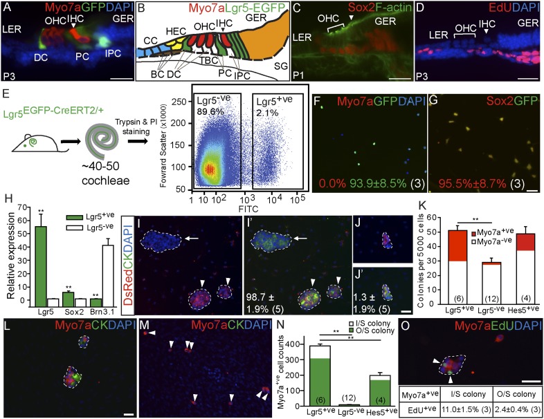
Inner ear hair cells are specialized sensory cells essential for auditory function. Previous studies have shown that the sensory epithelium is postmitotic, but it harbors cells that can behave as progenitor cells in vitro, including the ability to form new hair cells. Lgr5, a Wnt target gene, marks distinct supporting cell types in the neonatal cochlea. Here, we tested the hypothesis that Lgr5(+) cells are Wnt-responsive sensory precursor cells. In contrast to their quiescent in vivo behavior, Lgr5(+) cells isolated by flow cytometry from neonatal Lgr5(EGFP-CreERT2/+) mice proliferated and formed clonal colonies. After 10 d in culture, new sensory cells formed and displayed specific hair cell markers (myo7a, calretinin, parvalbumin, myo6) and stereocilia-like structures expressing F-actin and espin. In comparison with other supporting cells, Lgr5(+) cells were enriched precursors to myo7a(+) cells, most of which formed without mitotic division. Treatment with Wnt agonists increased proliferation and colony-formation capacity. Conversely, small-molecule inhibitors of Wnt signaling suppressed proliferation without compromising the myo7a(+) cells formed by direct differentiation. In vivo lineage tracing supported the idea that Lgr5(+) cells give rise to myo7a(+) hair cells in the neonatal Lgr5(EGFP-CreERT2/+) cochlea. In addition, overexpression of β-catenin initiated proliferation and led to transient expansion of Lgr5(+) cells within the cochlear sensory epithelium. These results suggest that Lgr5 marks sensory precursors and that Wnt signaling can promote their proliferation and provide mechanistic insights into Wnt-responsive progenitor cells during sensory organ development.


Conflict of interest statement

The authors declare no conflict of interest.

Figures

Fig. 1.
Fig. 1.
Lgr5+ cochlear supporting cells act as progenitor cells in vitro. (A) Cryosection of P3 Lgr5EGFP-CreERT2/+ cochlea showed GFP expression in the third Deiters’ cells (DC), inner pillar cells (PC), inner phalangeal cells (IPC), and the lateral greater epithelial ridge (GER). IHC, inner hair cells; LER, lesser epithelial ridge; OHC, outer hair cells. (B) Schematic depicting cell types in the P0–3 cochlea. BC, Boettcher cells; CC, Claudius cells; HEC, Hensen’s cells; SG, spiral ganglia; TBC, tympanic border cells. (C) Sox2 is expressed in Lgr5+ cells and other supporting cell types. (D) Cochlea from P3 mouse injected with EdU (P0–2) demonstrated no EdU uptake in the sensory epithelium. (E) Lgr5EGFP-CreERT2/+ cochleae were dissociated, and GFP+ and GFP cells were isolated using flow cytometry. (FH) Immunostaining and quantitative PCR showed that isolated GFP+ cells did not contain myo7a+ cells and robustly expressed Lgr5 and Sox2 but not brn3.1. (I and J) Lgr5+ cells isolated from Lgr5EGFP-CreERT2/+; Actin-DsRed mice were mixed (1:1) with those from Lgr5EGFP-CreERT2/+ mice and cultured. After 10 d, the majority of cytokeratin-positive colonies formed were monochromatic. I and J show DsRed and DAPI labeling only. I′ and J′ show DsRed, cytokeratin (CK), and DAPI labeling. (K) Lgr5+ and Hes5+ cells formed more colonies than Lgr5 cells. Forty percent of colonies from Lgr5+ cells contained myo7a+ cells. (LN) Lgr5+ cells generated more myo7a+ cells than Lgr5 or Hes5+ cells, most of which were outside (O/S) colonies. I/S, inside colonies. (O) In the presence of EdU, only a minority of myo7a+ cells were EdU+; double-positive cells were noted more commonly inside colonies. Data are represented as mean ± SD. **P < 0.01. In F, G, IK, N, and O, n is shown in parentheses. (Scale bars, 25 μm.)
Fig. 2.
Fig. 2.
Lineage tracing of Lgr5+ cells in the postnatal cochlea. (A and B) P3 Lgr5EGFP-CreERT2/+ cochlea showed GFP signals in supporting cell subtypes. GFP was rarely detected in myo7a+ cells and always was adjacent to a myo7a+, GFP+ cell at the third Deiters' cell position (arrowheads in B). (B′--B′′) Side views of reconstructed confocal images from B. B′ corresponds to the ′ position and B′′ to the ′′ position in B. (C) Tamoxifen was administered to P3 Lgr5EGFP-CreERT2/+; R26RtdTomato/+ mice, and cochleae were examined 2 and 6 d later. (D and E) Two days later, traced cells included the third Deiters' cells, inner pillar cells, inner phalangeal cells, and greater epithelial ridge cells. Traced GFP+, myo7a+ cells were rarely noted, and all were associated with myo7a+ cells at the the third Deiters' cell position (arrow in E′). (E′–E′′′′) Reconstructed z-stack images from E, showing merged labeling (E′), GFP and myo7a (E′′), tdTomato and myo7a (E′′′), and myo7a only (E′′′′). HC, hair cell. (F) Counts of GFP+ cells per cochlea. n is stated in parentheses. (G) At P9, traced cells included Hensen's cells, the second Deiters' cells (arrowheads), and hair cells (arrows). (H) GFP and myo7a labeling only. H′–H′′′′ are reconstructed z-stack images from G, showing merged labeling (H′), GFP and myo7a (H′′), tdTomato and myo7a (H′′′), and myo7a only (H′′′′). (I) Corn oil controls showed no tdTomato labeling. (J and K) Traced cells among outer and inner hair cells (OHC and IHC). (J′ and J′′) Side view and 3D reconstruction of confocal images from J′′. (K′–K′′′′) Reconstructed z-stack images from K, showing merged labeling (K′), GFP and myo7a (K′′), tdTomato and myo7a (K′′′), and myo7a only (K′′′′). (L) Counts of labeled hair cells and supporting cells from Lgr5EGFP-CreERT2/+; R26RtdTomato/+ mice. n is shown in parentheses. (Scale bars, 25 μm.)
Fig. 3.
Fig. 3.
Wnt signaling induces proliferation of Lgr5+ cells. (AC) Tamoxifen was given to P0–1 Lgr5EGFP-CreERT2/+; Catnbflox(exon3)/+ mice. Foci of GFP+ cells were noted 7 d later, abutting the inner hair cells and laterally in the lesser epithelial ridge. The medial foci (arrowheads) shifted adjacent to inner hair cells and disappeared by P21. (D) Pulse–chase experiments showed BrdU uptake (arrowheads) among formed foci. (D′ and D′′) High magnification and side view of foci. (E) Corn oil injection did not induce foci formation. (F) Foci counts per cochlea. (G and H) Tamoxifen was administered to P0–1 Lgr5EGFP-CreERT2/+; Catnbflox(exon3)/+; R26RtdTomato/+ mice. All GFP+ foci expressed tdTomato and Sox2 but not myo7a. G′–G′′′′ Side view of reconstructed images from G. (H′–H′′′′) Individual foci with tdTomato, GFP, Sox2, and Hoescht (H′), Sox2 only (H′′), tdTomato and Hoescht (H′′′), and GFP and Hoescht labeling (H′′′′). (IO) Purified Lgr5+ cells were cultured (10 d) with Wnt3a or R-spondin1. Treatment cells with Wnt3a or R-spondin1 increased cytokeratin-positive colonies, myo7a+ cells inside colonies, and EdU+, myo7a+ cells, but the total counts of myo7a+ cells were unaffected. *P < 0.05; **P < 0.01. In F and MO, n is shown in parentheses. (Scale bars, 25 μm in AE, G, H, and JL; 10 μm in H′–H′′′′.)
Fig. 4.
Fig. 4.
Wnt inhibition reduced proliferation of Lgr5+ cells in vitro. (A) Isolated Lgr5+ cells were cultured (10 d) with IWP-2 or aphidocilin. (BG) IWP-2 or aphidocilin decreased colony formation and EdU+ colonies. Drug treatment also reduced EdU+, myo7a+ cells and myo7a+ cells inside colonies but not outside colonies. In EG, n is shown in parentheses. **P < 0.01. (Scale bar, 25 μm in BD.) (H) Hypothetical model of Wnt-dependent proliferation and differentiation of Lgr5+ cells.

References

    1. Lin FR, Niparko JK, Ferrucci L. Hearing loss prevalence in the United States. Arch Intern Med. 2011;171:1851–1852. - PMC - PubMed
    1. Bermingham-McDonogh O, Reh TA. Regulated reprogramming in the regeneration of sensory receptor cells. Neuron. 2011;71:389–405. - PMC - PubMed
    1. Cruz RM, Lambert PR, Rubel EW. Light microscopic evidence of hair cell regeneration after gentamicin toxicity in chick cochlea. Arch Otolaryngol Head Neck Surg. 1987;113:1058–1062. - PubMed
    1. Corwin JT, Cotanche DA. Regeneration of sensory hair cells after acoustic trauma. Science. 1988;240:1772–1774. - PubMed
    1. Cafaro J, Lee GS, Stone JS. Atoh1 expression defines activated progenitors and differentiating hair cells during avian hair cell regeneration. Dev Dyn. 2007;236:156–170. - PubMed

Publication types

MeSH terms