. 2012 Jul;194(14):3651-60.
doi: 10.1128/JB.00114-12. Epub 2012 May 4.

Export requirements of pneumolysin in Streptococcus pneumoniae

Affiliations

Export requirements of pneumolysin in Streptococcus pneumoniae

Katherine E Price et al. J Bacteriol. 2012 Jul.

Abstract

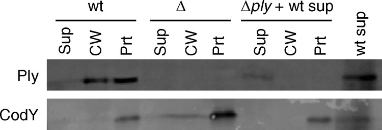
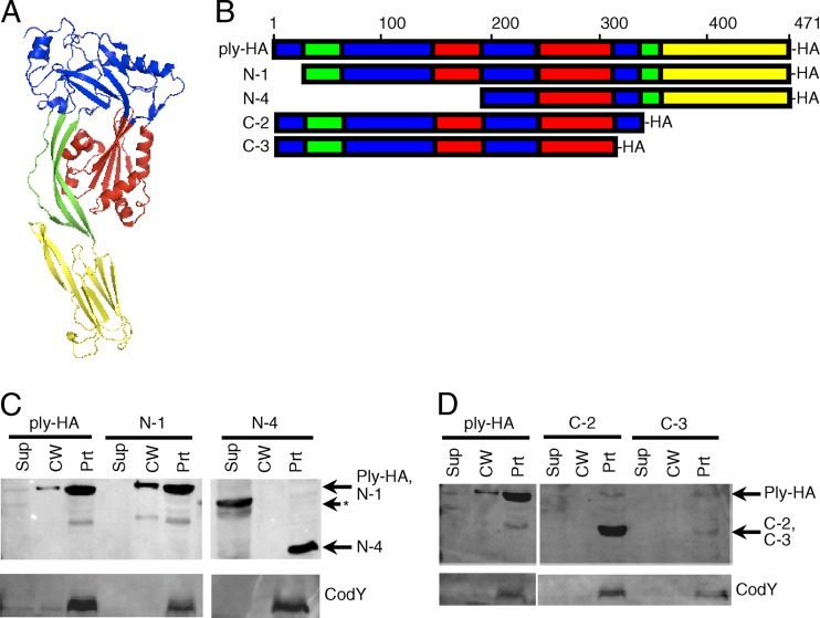
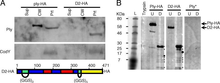
Streptococcus pneumoniae is a major causative agent of otitis media, pneumonia, bacteremia, and meningitis. Pneumolysin (Ply), a member of the cholesterol-dependent cytolysins (CDCs), is produced by virtually all clinical isolates of S. pneumoniae, and ply mutant strains are severely attenuated in mouse models of colonization and infection. In contrast to all other known members of the CDC family, Ply lacks a signal peptide for export outside the cell. Instead, Ply has been hypothesized to be released upon autolysis or, alternatively, via a nonautolytic mechanism that remains undefined. We show that an exogenously added signal sequence is not sufficient for Sec-dependent Ply secretion in S. pneumoniae but is sufficient in the surrogate host Bacillus subtilis. Previously, we showed that Ply is localized primarily to the cell wall compartment in the absence of detectable cell lysis. Here we show that Ply released by autolysis cannot reassociate with intact cells, suggesting that there is a Ply export mechanism that is coupled to cell wall localization of the protein. This putative export mechanism is capable of secreting a related CDC without its signal sequence. We show that B. subtilis can export Ply, suggesting that the export pathway is conserved. Finally, through truncation and domain swapping analyses, we show that export is dependent on domain 2 of Ply.


Figures

Fig 1
Fig 1
Ply released by autolysis cannot reassociate with intact cells. A wild-type (wt) culture was grown overnight and allowed to autolyze. The supernatant was collected, filtered, and added to a Δply culture. Wild-type and Δply cultures without added supernatant from an autolyzed culture were grown as positive and negative controls for the presence or absence of Ply, respectively. All cultures were grown to mid-exponential phase, fractionated into supernatant, cell wall, and protoplast compartments, and assayed for the presence of Ply and CodY by Western blotting. Equal cell equivalents were loaded on the gel. Sup, culture supernatant; CW, cell wall; Prt, protoplast.
Fig 2
Fig 2
SSRrgB-Ply fusion mRNA is made but protein is not detected in Streptococcus pneumoniae. (A) N-terminal DNA (top) and amino acid (bottom) sequences of SSRrgB-Ply. Text that is not in bold is Ply sequence. The RrgB signal peptide is in bold and underlined. The arrowhead indicates the predicted site of signal peptidase cleavage. (B) Western blot of cell wall fractionation of SSRrgB-Ply and acapsular TIGR4 cells. Cultures were grown to mid-exponential phase, fractionated into supernatant, cell wall, and protoplast compartments, and assayed for the presence of Ply and CodY by Western blotting. Equal cell equivalents of each fraction were loaded on the gel. A representative Western blot from two independent experiments is shown. Sup, culture supernatant; CW, cell wall; Prt, protoplast. (C) Quantitative RT-PCR on the ply transcript in acapsular TIGR4 and SSRrgB-Ply cells. Shown is the median fold change of the ply transcript of three replicates compared to acapsular TIGR4, normalized to gyrB; error bars indicate the range.
Fig 3
Fig 3
SSRrgB-Ply expression in Bacillus subtilis. (A) SSRrgB-Ply can be secreted when expressed in B. subtilis. SSRrgB-Ply was placed under the control of the Pspac* promoter in the amyE locus on the B. subtilis chromosome. All strains were grown to mid-exponential phase, fractionated into supernatant, cell wall, and protoplast compartments, and assayed for the presence of Ply and CodY by Western blotting. SSRrgB-Ply in B.s., SSRrgB-Ply fusion in B. subtilis; JH642, wild-type B. subtilis parent strain. Equal cell equivalents were loaded on the gel. Sup, culture supernatant; CW, cell wall; Prt, protoplast. (B) Hemolytic assay of culture supernatant, cell wall, and protoplast fractions of acapsular S. pneumoniae (acaps TIGR4), B. subtilis with ply (ply B.s.), B. subtilis with SS-ply (SS-ply B.s.), and B. subtilis parent strain JH642 (B.s). The ply in B. subtilis has the same distribution as in S. pneumoniae, with the highest hemolytic activity in the cell wall fraction. The supernatant fraction has the highest hemolytic activity in the SS-ply B. subtilis strain. No hemolytic activity was detected in the B. subtilis parent strain. The dotted line indicates the limit of detection. Bars show the means for three biological replicates; error bars indicate standard errors of the means (SEM).
Fig 4
Fig 4
Pfo expression in Streptococcus pneumoniae. (A) Amino acid alignment of Ply (top) and Pfo (bottom). Sequence homology does not begin until the third amino acid of Ply; therefore, fusions were made at this amino acid. The arrowhead indicates the site of signal peptidase cleavage in Pfo. The Pfo sequence not in bold was excluded in the Pfo signal sequence-less construct, thus deleting the Pfo signal peptide and six amino acids after the signal peptidase cleavage that share no homology to Ply. (B) Expression of signal sequence-less Pfo in S. pneumoniae. Signal sequence-less Pfo is expressed in S. pneumoniae, and a small amount localizes to the cell wall. The Pfo signal sequence-less allele (pfoNoSS) was used to replace ply at the native locus (Δply::pfoNoSS). This strain and a negative-control strain in which the ply deletion was replaced with a spectinomycin resistance gene (Δply::spc) were grown to mid-exponential phase, fractionated into supernatant, cell wall, and protoplast compartments, and assayed for the presence of Ply and CodY by Western blotting with anti-Pfo and anti-CodY antibodies. Equal cell equivalents were loaded on the gel.
Fig 5
Fig 5
Ply truncation mutants reveal that domain 2 of Ply may be required for export. (A) Model of Ply three-dimensional structure. The model was made with ModWeb software and manipulated with MacPyMol software. Domains are color coded: blue, domain 1; green, domain 2; red, domain 3; yellow, domain 4. (B) Schematic of primary sequences of Ply and truncation mutants. Domains are color coded as in panel A. Truncations of Ply were made from the N and C termini, tagged with an HA epitope at the C terminus of each construct, and crossed into the ply locus on the S. pneumoniae chromosome. Strains were grown to mid-exponential phase, fractionated into cell wall and protoplast compartments, and assayed for the presence of Ply and CodY by Western blotting. (C) N-terminal truncation mutants. The band in the supernatant lane for the N-4 truncation (shown by the asterisk) is of incorrect size and is unexplained. (D) C-terminal truncation mutants. Equal cell equivalents were loaded on the gel. Sup, culture supernatant; CW, cell wall; Prt, protoplast.
Fig 6
Fig 6
Ply with domain 2 replacement does not localize to the cell wall. Domain 2 was replaced with flexible linkers comprised of GGS repeats of appropriate length to maintain the three-dimensional spatial orientation of the remaining domains, tagged with a C-terminal HA epitope, and crossed into the ply locus on the S. pneumoniae chromosome. (A) Top, the ply-HA and D2 strains were grown to mid-exponential phase, fractionated into supernatant, cell wall, and protoplast compartments, and assayed for the presence of Ply and CodY by Western blotting. Equal cell equivalents were loaded on the gel. Sup, culture supernatant; CW, cell wall; Prt, protoplast. Bottom, schematic of the D2 replacement mutant, color coded by domain structure as in Fig. 5: blue, domain 1; green, domain 2; red, domain 3; yellow, domain 4. (B) Limited proteolysis of Ply with the domain 2 linker replacement reveals a wild-type conformation. Ply-HA and D2-HA were purified from strains Ply-HA and D2, respectively, via immunoprecipitation. His-tagged Ply toxoid protein (Ply*) served as a negative control for the anti-HA antibody. Approximately equivalent amounts of each purified protein were incubated either alone (U) or in the presence of identical amounts of trypsin (D). Trypsin alone was incubated under the same conditions. Equal amounts of each reaction product were analyzed for the presence of HA-tagged trypsin-cleaved peptides by Western blotting. The proteolytic degradation products of Ply-HA and D2-HA (marked with asterisks) are nearly identical. The presence of a single unique product in each (marked with circles) suggests a local change in surface accessibility that simultaneously exposes one trypsin cleavage site while deleting or masking another.
Fig 7
Fig 7
Ply domain 2 is necessary for export to the cell wall in S. pneumoniae. Domain 2 of Ply was replaced with domain 2 of Pfo and crossed into the ply locus on the S. pneumoniae chromosome. The strains carrying ply-HA and ply with Pfo's D2-HA were grown to mid-exponential phase, fractionated into supernatant, cell wall, and protoplast compartments, and assayed for the presence of Ply and CodY by Western blotting. Equal cell equivalents were loaded on the gel. Sup, culture supernatant; CW, cell wall; Prt, protoplast.
Fig 8
Fig 8
Ply localizes to the cell wall when expressed in Bacillus subtilis. The ply gene was placed under the control of the Pspac* promoter in the amyE locus on the B. subtilis chromosome (strain Ply B.s.). The B. subtilis parent strain, JH642, served as a control. Strains were grown to mid-exponential phase, fractionated into supernatant, cell wall, and protoplast compartments, and assayed for the presence of Ply and CodY by Western blotting. Equal equivalents were loaded on the gel. Sup, culture supernatant; CW, cell wall; Prt, protoplast.

Similar articles

Cited by

References

    1. Balachandran P, Hollingshead SK, Paton JC, Briles DE. 2001. The autolytic enzyme LytA of Streptococcus pneumoniae is not responsible for releasing pneumolysin. J. Bacteriol. 183:3108–3116 - PMC - PubMed
    1. Bassford P, Beckwith J. 1979. Escherichia coli mutants accumulating the precursor of a secreted protein in the cytoplasm. Nature 277:538–541 - PubMed
    1. Bassford PJ, JR, Silhavy TJ, Beckwith JR. 1979. Use of gene fusion to study secretion of maltose-binding protein into Escherichia coli periplasm. J. Bacteriol. 139:19–31 - PMC - PubMed
    1. Benton KA, Paton JC, Briles DE. 1997. Differences in virulence for mice among Streptococcus pneumoniae strains of capsular types 2, 3, 4, 5, and 6 are not attributable to differences in pneumolysin production. Infect. Immun. 65:1237–1244 - PMC - PubMed
    1. Bergmann S, Rohde M, Chhatwal GS, Hammerschmidt S. 2001. α-Enolase of Streptococcus pneumoniae is a plasmin(ogen)-binding protein displayed on the bacterial cell surface. Mol. Microbiol. 40:1273–1287 - PubMed

Publication types

LinkOut - more resources