. 2012 Nov;5(6):914-20.
doi: 10.1242/dmm.008995. Epub 2012 Apr 12.

Initiation of prostate cancer in mice by Tp53R270H: evidence for an alternative molecular progression

Affiliations

Initiation of prostate cancer in mice by Tp53R270H: evidence for an alternative molecular progression

Ruth L Vinall et al. Dis Model Mech. 2012 Nov.

Abstract

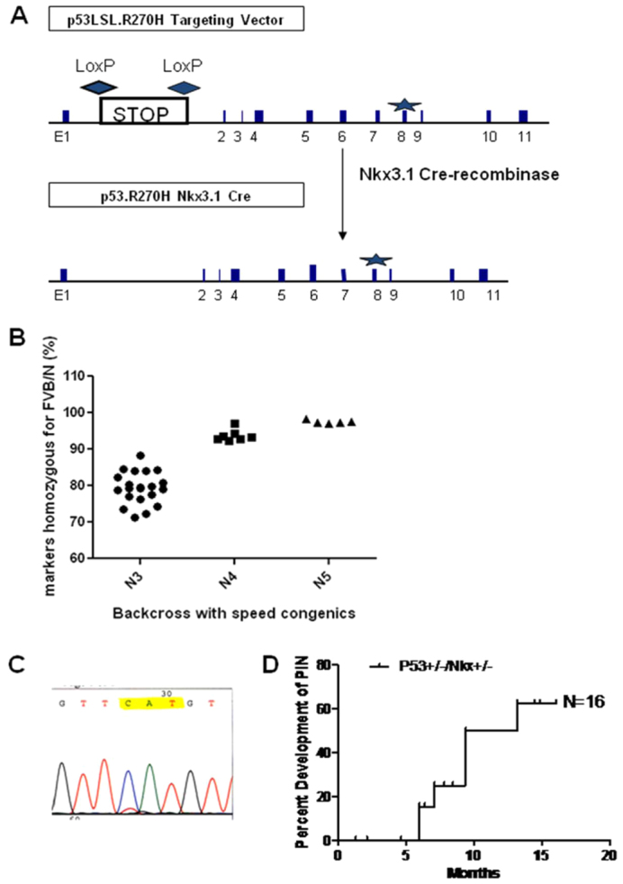
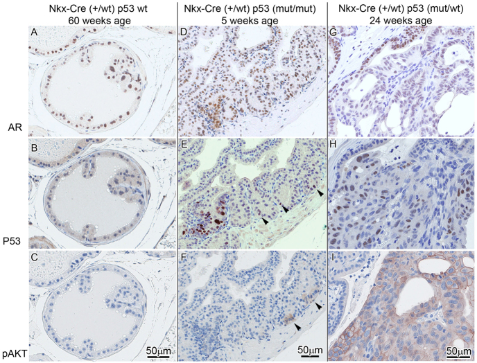
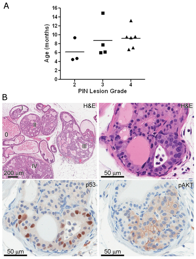
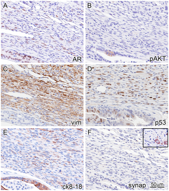
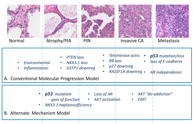
Tp53 mutations are common in human prostate cancer (CaP), occurring with a frequency of ∼30% and ∼70% in localized and metastatic disease, respectively. In vitro studies have determined several common mutations of Tp53 that have specific gain-of-function properties in addition to loss of function, including the ability to promote castration-resistant (CR) growth of CaP cells in some contexts. To date, a lack of suitable mouse models has prohibited investigation of the role played by Tp53 mutations in mediating CaP progression in vivo. Here, we describe the effects of conditional expression of a mutant Tp53 (Tp53(R270H); equivalent to the human hotspot mutant R273H) in the prostate epithelium of mice. Heterozygous "Tp53(LSL-R270H/+)" [129S4(Trp53(tm3Tyj))] and "Nkx3.1-Cre" [129S(Nkx3-1(tm3(cre)Mms))] mice with prostate-specific expression of the Tp53(R270H) mutation (p53(R270H/+) Nkx3.1-Cre mice) were bred onto an FVB/N background via speed congenesis to produce strain FVB.129S4(Trp53(tm3Tyj/wt)); FVB.129S(Nkx3-1(tm3(cre)Mms/wt)) and littermate genotype negative control mice. These mutant mice had significantly increased incidences of prostatic intraepithelial neoplasia (PIN) lesions, and these appeared earlier, compared with the Nkx3.1 haploinsufficient (Nkx3.1-Cre het) littermate mice, which did not express the Tp53 mutation. PIN lesions in these mice showed consistent progression and some developed into invasive adenocarcinoma with a high grade, sarcomatoid or epithelial-mesenchymal transition (EMT) phenotype. PIN lesions were similar to those seen in PTEN conditional knockout mice, with evidence of AKT activation concomitant with neoplastic proliferation. However, the invasive tumor phenotype is rarely seen in previously described mouse models of prostatic neoplasia. These data indicate that the Tp53(R270H) mutation plays a role in CaP initiation. This finding has not previously been reported. Further characterization of this model, particularly in a setting of androgen deprivation, should allow further insight into the mechanisms by which the Tp53(R270H) mutation mediates CaP progression.


Figures

Fig. 1.
Fig. 1.
Validation and characterization of the Tp53R270H/+Nkx3.1-Cre mouse. (A) Schematic representation of the targeting vector and the floxed allele following excision of the STOP cassette by Nkx3.1 Cre recombinase. The R270H mutation is located in exon 8 (marked as a star). (B) Congenic backcross onto FVB/NJ was performed to ensure a uniform strain background. Speed congenics (N3 through N5) produced a close to 100% FVB/NJ congenic mouse. (C) RT-PCR and sequencing analysis of laser capture microdissected PIN lesions confirmed that Nkx3.1-Cre-mediated deletion of the floxed STOP cassette resulted in prostate-specific expression of the p53 R270H mutation. (D) Well-developed PIN lesions were observed in Tp53R270H/+ Nkx3.1-Cre mice as early as 4 months and more than 60% of mice examined between 5 and 60 weeks developed grade 2 or higher PIN (n=16). Nkx3.1 haploinsufficient littermate mice (Nkx3.1-Cre; n=6), which did not express the Tp53 mutation, did not develop PIN even at 60 weeks (see Fig. 2).
Fig. 2.
Fig. 2.
Lesion progression in Tp53R270H/+ Nkx3.1-Cre mouse prostate. (A–C) In Tp53 wild-type mice with Nkx3.1 haploinsufficiency, foci of atypia were seen in older mice (60 weeks) but are not associated with well-developed PIN. p53 (B) and pAKT (C) are negative. (D–F) Early pre-PIN atypia/PIN 1 lesion in a 5-week-old mouse with p53 mutation/stabilization as detected by IHC (bottom left and arrowheads in E) seems to precede pAKT (focally seen arrowheads in F) and AR expression in normal and atypical areas (D). (G–I) At 24 weeks, areas with p53 mutation/stabilization in PIN 3–4 are accompanied by pAKT expression (I) and decreased AR nuclear intensity and percentage (G).
Fig. 3.
Fig. 3.
Distribution of PIN lesion grade among Tp53R270H/+ Nkx3.1-Cre mice. The scatterplot represents the highest grade lesion for a particular mouse (A), though often areas of lower grade were also seen (B). The horizontal line within each grade represents the mean age. Grading was performed as described previously (Park et al., 2002). Phenotypically, PIN lesions in Tp53R270H/+ Nkx3.1-Cre mice are similar to those seen in PTEN conditional knockout mice (B, upper right panel and lower 2 panels). Immunohistochemical analysis determined that p53 is stabilized in 10–70% of the cells within PIN lesions (B, lower left), further validating the expression of the p53 R270H mutation in these lesions. Levels of pAKT were elevated in almost all PIN lesions (B, lower right panel).
Fig. 4.
Fig. 4.
Immunophenotyping of invasive carcinoma reveals an EMT phenotype and AKT ‘de-addiction’. IHC (markers indicated lower right of each panel) on the same area (near-serial sections) of the periphery of an invasive tumor with an adjacent duct (bottom and lower left of each panel). This reveals strong AR expression in the duct, and weaker and lower percentage AR expression in the tumor cell nuclei (A). Focal pAKT is seen in the in situ atypia (B), corresponding to Tp53 stabilization/mutation (D). The tumor co-expresses vimentin (C) and luminal CK8/18 (E), and loses expression of pAKT (B). Although AKT activation is likely to be a driver of the PIN lesions, the invasive tumor seems to be independent of AKT (is ‘de-addicted’). The in situ areas are vimentin negative and cytokeratin positive (C,E). The tumor is negative for neuroendocrine differentiation as detected by a negative synaptophysin (F); inset is from the same tissue section showing a neural ganglion as internal positive control.
Fig. 5.
Fig. 5.
Heterogeneity of staining for AR in a PIN lesion and invasive carcinoma. In some PIN lesions (A), AR staining of the nuclei has become faint or is even lost (arrowheads) compared with nuclei of normal prostate epithelium (arrows). In invasive carcinoma, progressive loss of AR is seen with areas of strong nuclear staining in less than 50% of tumor cells (B), weak nuclear staining in less than 20% of cells (C; arrowheads) and areas of negative nuclear AR staining (D).
Fig. 6.
Fig. 6.
Schematic diagram of alternative molecular progression in prostate cancer compared with conventional model. CA, carcinoma; PIA, proliferative inflammatory atrophy.

References

    1. Acevedo V. D., Gangula R. D., Freeman K. W., Li R., Zhang Y., Wang F., Ayala G. E., Peterson L. E., Ittmann M., Spencer D. M. (2007). Inducible FGFR-1 activation leads to irreversible prostate adenocarcinoma and an epithelial-to-mesenchymal transition. Cancer Cell 12, 559–571 - PubMed
    1. Bauer J. J., Sesterhenn I. A., Mostofi F. K., McLeod D. G., Srivastava S., Moul J. W. (1996). Elevated levels of apoptosis regulator proteins p53 and bcl-2 are independent prognostic biomarkers in surgically treated clinically localized prostate cancer. J. Urol. 156, 1511–1516 - PubMed
    1. Bhatia-Gaur R., Donjacour A. A., Sciavolino P. J., Kim M., Desai N., Young P., Norton C. R., Gridley T., Cardiff R. D., Cunha G. R., et al. (1999). Roles for Nkx3.1 in prostate development and cancer. Genes Dev. 13, 966–977 - PMC - PubMed
    1. Byrne R. L., Horne C. H., Robinson M. C., Autzen P., Apakama I., Bishop R. I., Neal D. E., Hamdy F. C. (1997). The expression of waf-1, p53 and bcl-2 in prostatic adenocarcinoma. Br. J. Urol. 79, 190–195 - PubMed
    1. Chi S. G., deVere White R. W., Meyers F. J., Siders D. B., Lee F., Gumerlock P. H. (1994). p53 in prostate cancer: frequent expressed transition mutations. J. Natl. Cancer Inst. 86, 926–933 - PubMed

Publication types

MeSH terms