Comparative Study
. 2012 Jun 29;287(27):23024-33.
doi: 10.1074/jbc.M112.367011. Epub 2012 May 4.

Exercise is more effective than diet control in preventing high fat diet-induced β-amyloid deposition and memory deficit in amyloid precursor protein transgenic mice

Affiliations
Comparative Study

Exercise is more effective than diet control in preventing high fat diet-induced β-amyloid deposition and memory deficit in amyloid precursor protein transgenic mice

Masato Maesako et al. J Biol Chem. .

Abstract

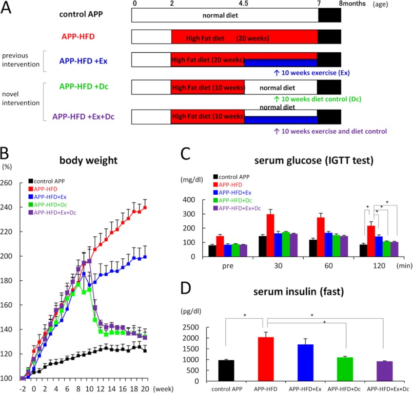
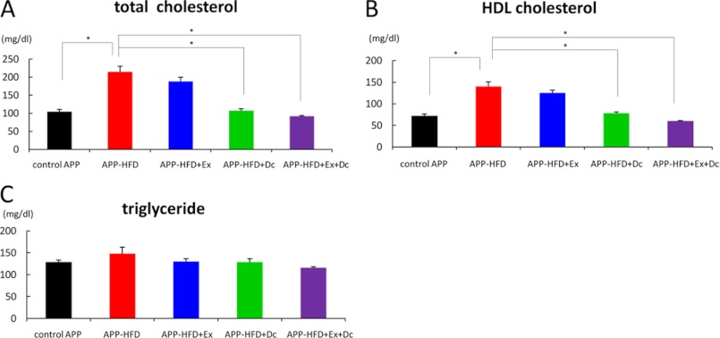
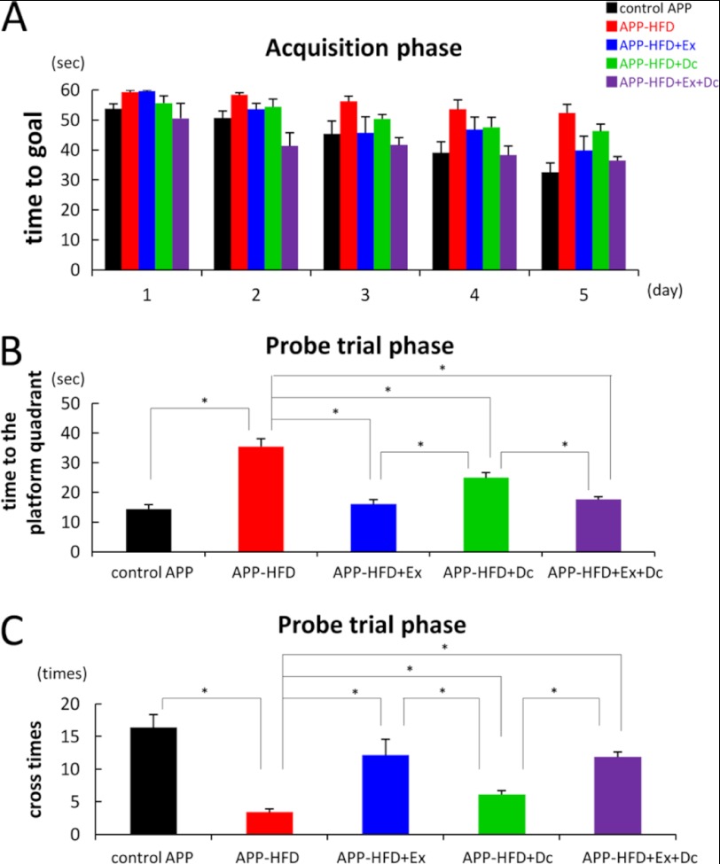
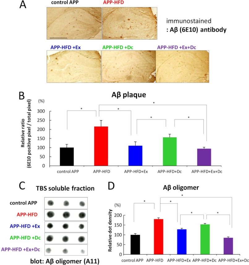
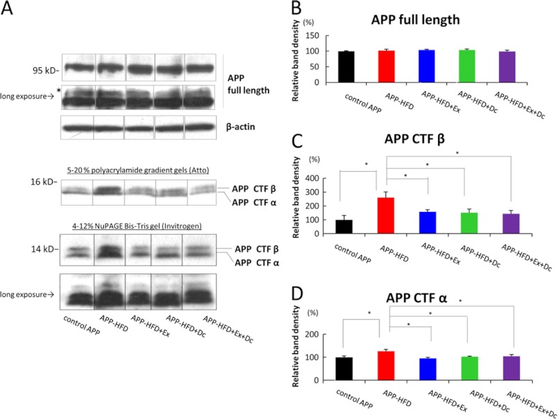
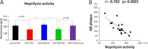
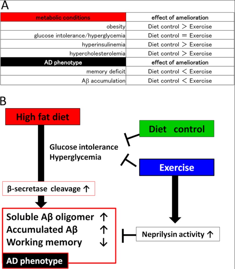
Accumulating evidence suggests that some dietary patterns, specifically high fat diet (HFD), increase the risk of developing sporadic Alzheimer disease (AD). Thus, interventions targeting HFD-induced metabolic dysfunctions may be effective in preventing the development of AD. We previously demonstrated that amyloid precursor protein (APP)-overexpressing transgenic mice fed HFD showed worsening of cognitive function when compared with control APP mice on normal diet. Moreover, we reported that voluntary exercise ameliorates HFD-induced memory impairment and β-amyloid (Aβ) deposition. In the present study, we conducted diet control to ameliorate the metabolic abnormality caused by HFD on APP transgenic mice and compared the effect of diet control on cognitive function with that of voluntary exercise as well as that of combined (diet control plus exercise) treatment. Surprisingly, we found that exercise was more effective than diet control, although both exercise and diet control ameliorated HFD-induced memory deficit and Aβ deposition. The production of Aβ was not different between the exercise- and the diet control-treated mice. On the other hand, exercise specifically strengthened the activity of neprilysin, the Aβ-degrading enzyme, the level of which was significantly correlated with that of deposited Aβ in our mice. Notably, the effect of the combination treatment (exercise and diet control) on memory and amyloid pathology was not significantly different from that of exercise alone. These studies provide solid evidence that exercise is a useful intervention to rescue HFD-induced aggravation of cognitive decline in transgenic model mice of AD.


Figures

FIGURE 1.
FIGURE 1.
Diet control ameliorated HFD-induced obesity and diabetic conditions when compared with exercise. A, schematic presentation of the interventions targeting metabolic conditions. As described previously (7), APPSwe/Ind mice were maintained with a standard diet in standard laboratory cages until they were 2–3 months old. Then, age- and sex-matched mice were separated into five groups. In the control group, the mice were fed with a standard diet in standard laboratory cages for 20 weeks (control APP mice) (top row, n = 9). In the HFD-induced group, mice were fed HFD in standard laboratory cages for 20 weeks (APP-HFD mice) (second row, n = 10). In the HFD with exercise-induced group, mice spent 10 weeks in standard laboratory cages and then spent 10 weeks in enrichment cages with HFD (APP-HFD+Ex mice) (third row, n = 8). As a novel intervention, in the diet-control-induced group, after 10 weeks of HFD, we used a standard diet for another 10 weeks (APP-HFD+Dc mice) (fourth row, n = 7). In the combination group with exercise plus diet control, mice spent 10 weeks in standard laboratory cages with HFD and then spent 10 weeks in enrichment cages with a standard diet (APP-HFD+Ex+Dc mice) (bottom row, n = 7). After 20 weeks, metabolic conditions of these mice were analyzed followed by ethological, histochemical, and biochemical analyses targeting AD pathophysiology. B, relative body weight changes over 20 weeks. The body weight 2 weeks before each diet was regarded as the baseline (100%). Diet control and its combination with exercise significantly inhibited the HFD-induced increase of body weight. C, blood glucose levels during glucose tolerance test after an intraperitoneal injection of glucose (2 g/kg of body weight). The fasting glucose level (pre) and glucose tolerance in APP-HFD+Dc mice (F(4,159) = 26.49, *, p < 0. 001) and APP-HFD+Ex+Dc mice (*, p < 0.001) were clearly improved. D, serum insulin levels during fasting. The serum insulin levels in APP-HFD+Dc mice (F(4, 36) = 9.3, *, p = 0.003) and APP-HFD+Ex+Dc mice (*, p < 0.001) were significantly decreased when compared with that in APP-HFD mice.
FIGURE 2.
FIGURE 2.
Diet control ameliorated HFD-induced lipid dysfunction when compared with exercise. A, plasma total cholesterol levels. The total cholesterol levels in APP-HFD+Dc mice (F(4, 36) = 30.29, *, p < 0.001) and APP-HFD+Ex+Dc mice (*, p < 0.001) were significantly decreased when compared with the level in APP-HFD mice. B, plasma HDL cholesterol levels. The HDL cholesterol levels in APP-HFD+Dc mice (F(4, 36) = 30.96, *, p < 0.001) and APP-HFD+Ex+Dc mice (*, p < 0.001) were significantly decreased when compared with the level in APP-HFD mice. C, plasma triglyceride levels. There was no difference among control, APP-HFD, APP-HFD+Ex, APP-HFD+Dc, and APP-HFD+Ex+Dc mice (F(4, 36) = 1.65, not significant).
FIGURE 3.
FIGURE 3.
Exercise ameliorated HFD-induced memory deficit when compared with diet control. A, escape latency in the acquisition phase. APP-HFD+Ex mice took a shorter time to the platform than APP-HFD+Dc mice. B, the time to the target quadrant in the probe trial phase. APP-HFD+Ex mice took a shorter time to the platform than APP-HFD+Dc mice (F(4, 36) = 23. 03, *, p = 0. 041). C, the number of entries into the target quadrant in the probe trial phase. APP-HFD+Dc mice were significantly impaired in the number of times they crossed the platform when compared with APP-HFD+Ex mice (F(4, 36) = 13.59, *, p = 0. 013).
FIGURE 4.
FIGURE 4.
Exercise ameliorated HFD-induced Aβ accumulation when compared with diet control. A, immunohistochemical analysis using anti-Aβ (6E10) antibody. Representative images of Aβ-immunostained hippocampus sections from control APP, APP-HFD, APP-HFD+Ex, APP-HFD+Dc, and APP-HFD+Ex+Dc induced mice, respectively, are shown. Scale bar, 2 mm. B, cerebral Aβ loads determined by immunohistochemical and morphometric analyses. The cerebral Aβ deposition was significantly decreased in APP-HFD+Ex mice when compared with that in APP-HFD+Dc mice (F(4, 15) = 18.35, *, p = 0. 039). C, the amount of Aβ oligomers in the TBS-soluble fractions of control-APP, APP-HFD, APP-HFD+Ex, APP-HFD+Dc, and APP-HFD+Ex+Dc mice analyzed by filter trap assay using anti-Aβ oligomer (A11) antibody. As described in Ref. , the Aβ monomer and low weight oligomers passed through the membrane pore (200-nm pore size), and high weight oligomers were detected in this assay. D, statistical analysis of dot density. The average band density of the control APP samples was regarded as 100%, and that of other groups was relatively indicated. The relative density of APP-HFD+Ex mice was significantly decreased when compared with that of APP-HFD+Dc mice (F(4, 10) = 47.42, *, p = 0.011).
FIGURE 5.
FIGURE 5.
Both diet control and exercise reduced APP-CTFβ accumulation. A, immunoblotting analysis of full-length APP and APP-CTFα and APP-CTFβ. Full-length APP and APP CTFs (CTFα and CTFβ) were detected by anti-APP C terminus antibody. β-Actin was detected as loading control. Long exposure indicated that the same film was exposed for a longer time. The asterisk indicates glycosylated full-length APP. To analyze APP CTFs in detail, two kinds of gels (5–20% polyacrylamide gradient gels and 4–12% NuPAGE Bis-Tris gels) were used. Unfortunately, we could not clarify the mobility of CTFs bands caused by phosphorylation presumably because of the gel conditions of our experiment. B, statistical analysis of full-length APP. The band of full-length APP was normalized by that of β-actin. The band density of the control was regarded as 100%, and that of other groups was relatively indicated. There was no statistically significant difference among control APP, APP-HFD, APP-HFD+Ex, APP-HFD+Dc, and APP-HFD+Ex+Dc mice (F(4, 10) = 0.47, not significant). C, statistical analysis of APP-CTFβ. The band of APP-CTFβ was normalized by that of full-length APP. The band density of APP-CTFβ in APP-HFD mice was increased when compared with that in control APP mice. However, the band densities of APP-CTFβ in APP-HFD+Ex (F(4, 10) = 4.27, *, p = 0.003), APP-HFD+Dc (*, p = 0.021), and APP-HFD+Ex+Dc (*, p = 0.023) mice were significantly decreased when compared with that in APP-HFD mice. There was no difference between APP-CTFβ in APP-HFD+Ex mice and that in APP-HFD+Dc mice. D, statistical analysis of APP-CTFα. The band of APP-CTFα was normalized by that of full-length APP. The band density of APP-CTFα in APP-HFD mice was increased when compared with that in control APP mice. The band densities of APP-CTFα in APP-HFD+Ex (F(4, 10) = 4.36, *, p = 0.034), APP-HFD+Dc (*, p = 0.014), and APP-HFD+Ex+Dc (*, p = 0.024) mice were significantly decreased when compared with that in APP-HFD mice. There was no difference between APP-CTFα in APP-HFD+Ex mice and that in APP-HFD+Dc mice.
FIGURE 6.
FIGURE 6.
Exercise specifically rescued HFD-induced deterioration of neprilysin activity. A, In vitro enzyme activity assay of neprilysin using the fluorescence substrate. The activity of neprilysin in APP-HFD mice tends to be decreased when compared with that in control APP mice (F(4, 15) = 5.58, p = 0.061). On the other hand, neprilysin activity in APP-HFD+Ex mice was significantly higher than that in APP-HFD mice (*, p = 0.023) or that in APP-HFD+Dc mice (*, p = 0.032). B, significant correlation was established by comparing the activity of neprilysin and the level of cerebral Aβ deposition, using Pearson's correlation coefficients. The activity of neprilysin was negatively correlated with the level of accumulated Aβ (r = −0.782, *, p = 0.00003).
FIGURE 7.
FIGURE 7.
Schematic presentation of our study. A, the classification of the results in the present study. The items we analyzed in this study are included in the left column, whereas the effect of amelioration is showed in the right column. As shown in this table, Diet control > Exercise indicated that diet control ameliorated better than exercise. Diet control significantly improved HFD-induced metabolic conditions, including obesity, hyperinsulinemia, and hypercholesterolemia, better than exercise. However, exercise decreased soluble Aβ oligomers as well as deposited Aβ and ameliorated memory impairment better than diet control. B, schematic presentation of our hypothesis: how diet control or exercise ameliorated HFD-induced memory deficits and Aβ accumulation. HFD leads to glucose intolerance and hyperglycemia, which may lead to the up-regulation of β-secretase activity. This up-regulation increases soluble Aβ oligomers as well as deposited Aβ levels followed by memory deficit (7). On the other hand, both diet control and exercise ameliorate HFD-induced glucose intolerance and hyperglycemia, thereby decreasing soluble Aβ oligomer and fibrillar Aβ levels by inhibiting Aβ production. However, exercise specifically strengthens the enzymatic activity of neprilysin, which degrades Aβ in the brain.

References

    1. Finder V. H. (2010) Naturally secreted oligomers of amyloid-β protein potently inhibit hippocampal long-term potentiation in vivo. J. Alzheimers Dis. 22, Suppl. 3, 5–19 - PubMed
    1. Walsh D. M., Klyubin I., Fadeeva J. V., Cullen W. K., Anwyl R., Wolfe M. S., Rowan M. J., Selkoe D. J. (2002) Naturally secreted oligomers of amyloid-β protein potently inhibit hippocampal long-term potentiation in vivo. Nature 416, 535–539 - PubMed
    1. Lesné S., Koh M. T., Kotilinek L., Kayed R., Glabe C. G., Yang A., Gallagher M., Ashe K. H. (2006) A specific amyloid-β protein assembly in the brain impairs memory. Nature 440, 352–357 - PubMed
    1. Shankar G. M., Li S., Mehta T. H., Garcia-Munoz A., Shepardson N. E., Smith I., Brett F. M., Farrell M. A., Rowan M. J., Lemere C. A., Regan C. M., Walsh D. M., Sabatini B. L., Selkoe D. J. (2008) Amyloid-β protein dimers isolated directly from Alzheimer brains impair synaptic plasticity and memory. Nat. Med. 14, 837–842 - PMC - PubMed
    1. Luchsinger J. A., Tang M. X., Shea S., Mayeux R. (2002) Caloric intake and the risk of Alzheimer disease. Arch. Neurol. 59, 1258–1263 - PubMed

Publication types

MeSH terms