. 2012 Jun 29;287(27):23079-94.
doi: 10.1074/jbc.M111.328757. Epub 2012 May 4.

Requirements for stress granule recruitment of fused in sarcoma (FUS) and TAR DNA-binding protein of 43 kDa (TDP-43)

Affiliations

Requirements for stress granule recruitment of fused in sarcoma (FUS) and TAR DNA-binding protein of 43 kDa (TDP-43)

Eva Bentmann et al. J Biol Chem. .

Abstract

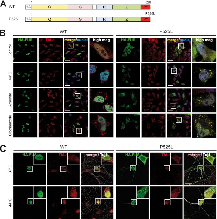
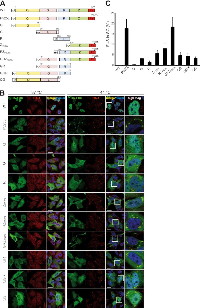
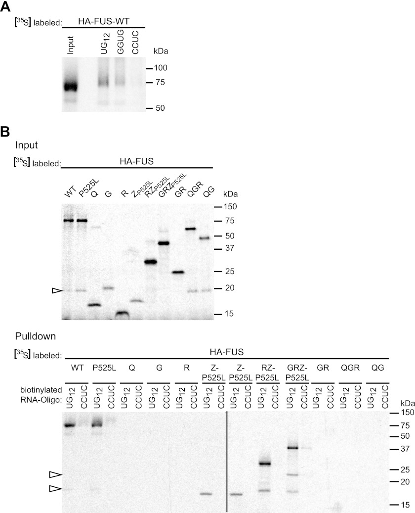
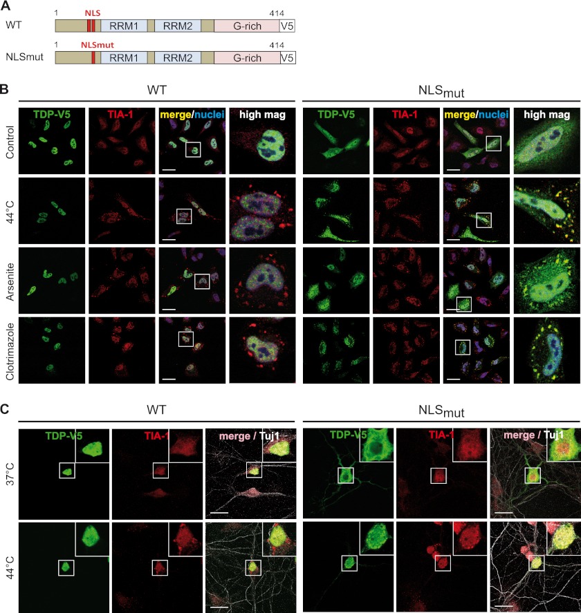
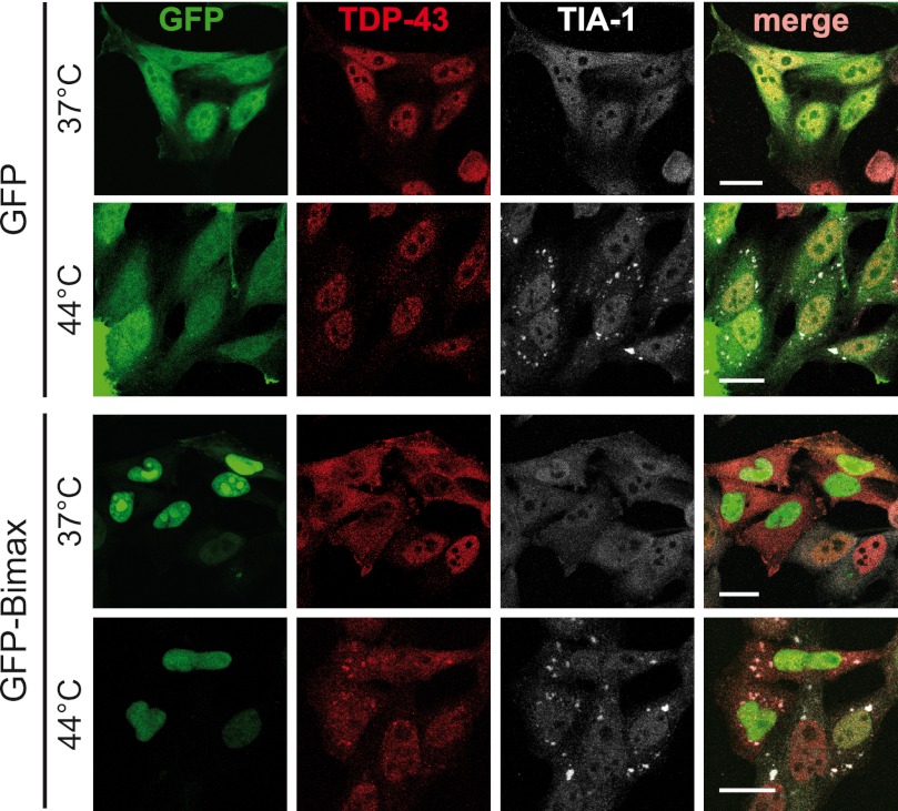
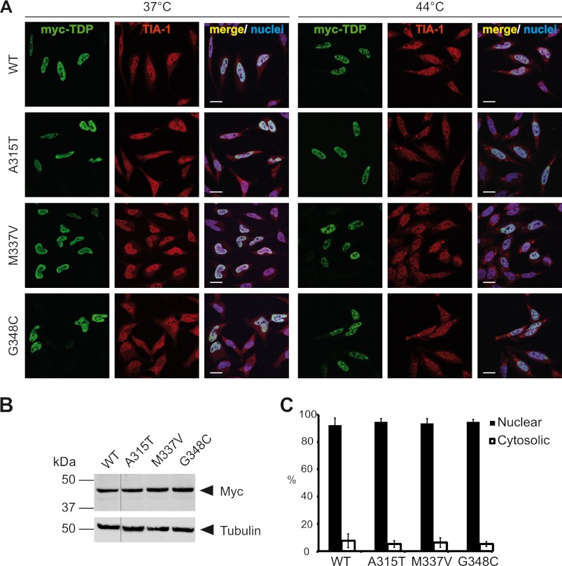
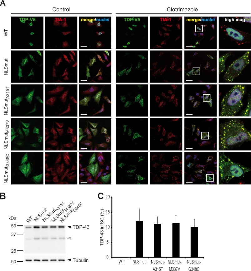
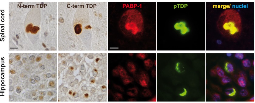
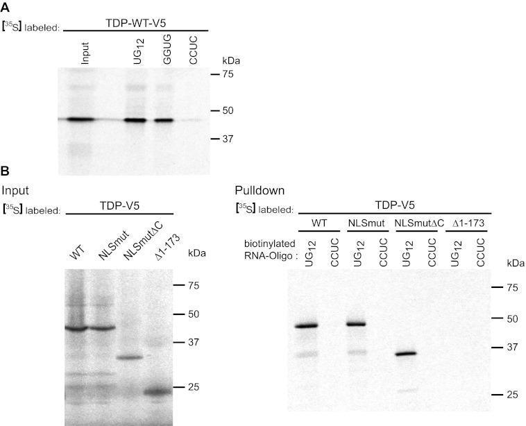
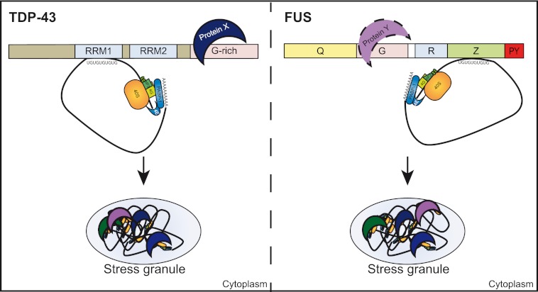
Cytoplasmic inclusions containing TAR DNA-binding protein of 43 kDa (TDP-43) or Fused in sarcoma (FUS) are a hallmark of amyotrophic lateral sclerosis (ALS) and several subtypes of frontotemporal lobar degeneration (FTLD). FUS-positive inclusions in FTLD and ALS patients are consistently co-labeled with stress granule (SG) marker proteins. Whether TDP-43 inclusions contain SG markers is currently still debated. We determined the requirements for SG recruitment of FUS and TDP-43 and found that cytoplasmic mislocalization is a common prerequisite for SG recruitment of FUS and TDP-43. For FUS, the arginine-glycine-glycine zinc finger domain, which is the protein's main RNA binding domain, is most important for SG recruitment, whereas the glycine-rich domain and RNA recognition motif (RRM) domain have a minor contribution and the glutamine-rich domain is dispensable. For TDP-43, both the RRM1 and the C-terminal glycine-rich domain are required for SG localization. ALS-associated point mutations located in the glycine-rich domain of TDP-43 do not affect SG recruitment. Interestingly, a 25-kDa C-terminal fragment of TDP-43, which is enriched in FTLD/ALS cortical inclusions but not spinal cord inclusions, fails to be recruited into SG. Consistently, inclusions in the cortex of FTLD patients, which are enriched for C-terminal fragments, are not co-labeled with the SG marker poly(A)-binding protein 1 (PABP-1), whereas inclusions in spinal cord, which contain full-length TDP-43, are frequently positive for this marker protein.


Figures

FIGURE 1.
FIGURE 1.
Cytosolic FUS is recruited to SG upon treatment with various stressors. A, shown is a schematic diagram of FUS wild-type (WT) and P525L mutant used for transient transfection in HeLa cells. HA, HA epitope tag; Q, glutamine-rich domain; G, glycine-rich domain; R, RRM domain; Z, arginine-glycine-glycine (RGG) zinc finger domain. B, HeLa cells were transiently transfected with N-terminal-HA-tagged FUS-WT or FUS-P525L. 24 h after transfection cells were subjected to heat shock (44 °C for 1 h), sodium arsenite (0.5 mm for 30 min), or clotrimazole (20 μm for 30 min) or were left untreated (Control). Cells were fixed, stained with an HA-specific antibody (green), a TIA-1-specific antibody (red), and a nuclear counterstain (blue) and analyzed by confocal microscopy. Panels to the right show a higher magnification of the boxed region. Although FUS-WT remained nuclear, FUS-P525L was sequestered into SG under all stress conditions examined. Scale bars, 20 μm. C, primary rat hippocampal neurons were transiently transfected with HA-FUS-WT or P525L on DIV 7. 48 h after transfection, neurons were subjected to heat shock (44 °C) for 1 h or left untreated (37 °C). Neurons were fixed and stained with an HA-specific antibody (green), a TIA-1-specific antibody (red), and the neuronal marker antibody Tuj1 (white) to visualize neuronal morphology. FUS-P525L showed cytoplasmic mislocalization and was recruited to TIA-1-positive SG upon heat stress. Insets in the upper right corner show a higher magnification of the boxed region. Scale bars, 20 μm.
FIGURE 2.
FIGURE 2.
The C-terminal RGG-zinc finger domain of FUS is the most important domain for SG recruitment. A, shown is a schematic diagram of different FUS constructs analyzed for SG recruitment. The P525L mutation was introduced into the PY-NLS to obtain proteins mislocalized in the cytosol. B, shown is immunocytochemistry of HeLa cells expressing the different FUS constructs shown in A. Before fixation, cells were subjected to heat shock (44 °C for 1 h) or left untreated (37 °C). Cells were stained with an HA-specific antibody (green), a TIA-1-specific antibody (red), and a nuclear counterstain (blue) and analyzed by confocal microscopy. Panels to the right show a higher magnification of the boxed region. The Z domain is most important for SG recruitment, whereas the Q domain is dispensable. The G and R domains also contribute to SG recruitment but to a lesser extent than Z. Scale bars = 20 μm. C, the percentage of FUS localized in TIA-1-positive SG was quantified using ImageJ. 10–20 cells were analyzed in a blinded manner, means across all cells were calculated, and S.D. are indicated by error bars. Note that the percentage of FUS-P525L in SG seems surprisingly low when looking at the corresponding confocal images in B. However, SG are very small compared with the remaining cellular volume, and therefore, FUS-P525L diffusely distributed in the cytosol and nucleus amounts to a significant percentage of the total protein (more than 80%).
FIGURE 3.
FIGURE 3.
The RGG-zinc finger domain of FUS binds to UG-rich RNA. A, FUS-WT was in vitro translated in the presence of [35S]methionine (left lane, input) and was analyzed for binding to different RNA oligonucleotides immobilized on streptavidin beads (right lanes, UG12, UGUGUGUGUGUGUGUGUGUGUGUG; GGUG, UUGUAUUUUGAGCUAGUUUGGUGAU; CCUC, UUGUAUUUUGAGCUAGUUUCCUCAU). FUS was pulled down most efficiently by UG12 and to a lesser extent by GGUG RNA. B, the indicated FUS constructs were in vitro translated in the presence of [35S]methionine (upper panel, Input). Biotinylated UG12 RNA immobilized on streptavidin beads was used to pull down radioactively labeled proteins (lower panel, Pulldown). CCUC RNA was used as a negative control. FUS-WT and P525L and all proteins comprising the ZP525L domain were specifically pulled down by UG12 RNA, whereas the other proteins did not show detectable RNA binding. Open arrowheads indicate degradation products.
FIGURE 4.
FIGURE 4.
Cytosolic mislocalization is a prerequisite for SG recruitment of TDP-43. A, shown is a schematic diagram of TDP-43 wild-type (WT) and NLS mutant (NLSmut). NLSmut, triple point mutation in the classical nuclear localization signal (K83A/R84A/K85A); G-rich, glycine-rich domain; V5, V5 epitope tag. B, C-terminal-V5-tagged TDP-WT or NLSmut were transiently transfected into HeLa cells and 24 h later were subjected to heat shock (44 °C for 1 h), sodium arsenite (0.5 mm for 30 min), or clotrimazole (20 μm for 30 min) treatment or were left untreated (Control). Cells were fixed, stained with a V5 (green) and TIA-1 (red)-specific antibody and a nuclear counterstain (blue), and analyzed by confocal microscopy. Panels to the right show a higher magnification of the boxed region. Although the cytosolic NLS mutant was sequestered into SG, TDP-WT remained nuclear under all stress conditions examined. Scale bars = 20 μm. C, primary rat hippocampal neurons were transiently transfected with V5-tagged TDP-WT or NLSmut. 48 h post-transfection, neurons were subjected to heat shock (44 °C) for 1 h or left untreated (37 °C). Neurons were fixed and stained with a V5-specific antibody (green), a TIA-1-specific antibody (red), and the neuronal marker antibody Tuj1 (white) to visualize neuronal morphology. NLSmut showed partial cytoplasmic mislocalization and was recruited to TIA-1-positive SG upon heat stress. Insets in the upper right corner show a higher magnification of the boxed region. Scale bars, 20 μm.
FIGURE 5.
FIGURE 5.
Endogenous TDP-43 is sequestered into heat shock-induced SG upon inhibition of Importin α/β-dependent nuclear import. HeLa cells were transfected with an Importin α/β-specific peptide inhibitor fused to GFP (GFP-Bimax) or GFP as a control (green). 24 h post-transfection cells were subjected to heat shock (44 °C for 1 h) or kept at control temperature (37 °C) before fixation. Cells were co-stained for endogenous TDP-43 (red) and TIA-1 (white) and analyzed by confocal microscopy. Expression of GFP-Bimax resulted in cytosolic mislocalization of endogenous TDP-43 and recruitment into SG upon heat shock. Under control conditions (GFP), TDP-43 was predominantly nuclear and did not colocalize with SG after heat shock. Scale bars = 20 μm.
FIGURE 6.
FIGURE 6.
ALS-associated TARDBP mutations do not alter the cellular localization of TDP-43. A, Myc-tagged wild-type TDP-WT or TDP-43 carrying the indicated ALS-associated point mutations (A315T, M337V, or G348C) were transiently transfected into HeLa cells. 24 h post-transfection cells were subjected to heat shock (44 °C for 1 h) or kept at control temperature (37 °C). Afterward cells were fixed, stained with a myc (green) and TIA-1 (red)-specific antibody and a nuclei counterstain (blue), and analyzed by confocal microscopy. Both TDP-WT and the ALS-associated point mutants were nuclear under control conditions and remained nuclear upon heat shock. Scale bars = 20 μm. B, shown are expression levels of TDP-43 constructs used in A. Total cell lysates were analyzed by immunoblotting with a myc-specific antibody (upper panel). Tubulin served as a loading control (lower panel). All lanes were from the same exposure of the same blot. C, quantification of nuclear and cytosolic fluorescence intensities of myc staining at 37 °C is shown. Error bars indicate S.D.
FIGURE 7.
FIGURE 7.
ALS-associated TARDBP mutations do not affect SG recruitment of cytosolic TDP-43. ALS-associated point mutations (A315T, M337V, G348C) were introduced into the TDP-43 NLS mutant (NLSmutA315T, NLSmutM337V, NLSmutG348C), and the effect of mutations on SG recruitment was analyzed. A, HeLa cells transiently transfected with the indicated TDP-43 constructs were incubated with clotrimazole for 30 min or left untreated (Control). Cells were fixed, stained with a V5 (green) and TIA-1 (red)-specific antibody and a nuclear counterstain (blue), and analyzed by confocal microscopy. ALS-associated point mutations did not affect SG recruitment of cytosolic TDP-43. Scale bars = 20 μm. B, protein levels in total cell lysates were analyzed by immunoblotting with a V5-specific antibody (upper panel), and tubulin served as a loading control (lower panel). The black arrowhead indicates full-length TDP-43, and the white arrowhead indicates caspase-generated 35-kDa CTF frequently observed under transient transfection conditions (61, 75). C, the percentage of TDP-43 localized in TIA-1-positive SG was quantified using ImageJ. 15–20 cells were analyzed in a blinded manner, means across all cells were calculated, and S.D. are indicated by error bars.
FIGURE 8.
FIGURE 8.
TDP-43 inclusions in spinal cord but not hippocampus are frequently co-labeled for the SG marker protein PABP-1. TDP-43 immunohistochemistry performed on formalin-fixed, paraffin-embedded tissue sections of spinal cord (upper panels) or hippocampus (lower panels) from FTLD-TDP and ALS-TDP cases is shown. Staining with N-terminal and C-terminal TDP-43-specific antibodies demonstrated labeling of neuronal cytoplasmic inclusions in motor neurons in the spinal cord with both antibodies (ALS case #1 shown), whereas inclusions in dentate granule neurons in the hippocampus were labeled only with the C-terminal antibody (FTLD-TDP case #1 shown). Double-label immunofluorescence stainings of the same cases showed co-labeling of phospho-TDP-43 positive inclusions (green) with the SG marker protein PABP-1 (red) in the spinal cord but not in cortical inclusions. Nuclei were stained with DAPI (blue). Scale bars = 10 μm.
FIGURE 9.
FIGURE 9.
25 kDa C-terminal fragment and a C-terminal deletion mutant of TDP-43 are poorly sequestered into SG. A, shown is a schematic diagram of TDP-43 deletion mutants analyzed for SG recruitment. The deletion mutant Δ1–173 was chosen to mimic the 25-kDa CTF found to be deposited in cortical regions of FTLD-TDP patients (4, 59). The C-terminal deletion mutant (NLSmutΔC) lacks the prion-like glycine-rich domain. B, the indicated TDP-43 constructs were transiently transfected in HeLa cells. Before fixation, cells were treated with clotrimazole (20 μm, 30 min) or left untreated (control). Subsequently, cells were stained with a V5 (green) and TIA-1 (red)-specific antibody and a nuclear counterstain (blue) and analyzed by confocal microscopy. Panels to the right show a higher magnification of the boxed region. In contrast to full-length TDP-NLSmut, both deletion mutants remained predominantly diffuse in the cytosol upon heat shock and were poorly recruited to SG. Scale bars = 20 μm. C, the percentage of TDP-43 localized in TIA-1-positive SG was quantified using ImageJ. 15–20 cells were analyzed in a blinded manner, means across all cells were calculated, and S.D. are indicated by error bars. D, protein levels in total cell lysates were analyzed by immunoblotting with a V5-specific antibody (upper panel); tubulin served as a loading control (lower panel). Black arrowheads indicate full-length TDP-NLSmut or the two deletion mutants, and white arrowheads indicate degradation products. All lanes were from the same exposure of the same blot.
FIGURE 10.
FIGURE 10.
The C-terminal deletion mutant of TDP-43 still binds to UG12 RNA. A, TDP-WT was in vitro translated in the presence of [35S]methionine (left lane, Input) and was analyzed for binding to different RNA oligonucleotides immobilized on streptavidin beads (right lanes, UG12, UGUGUGUGUGUGUGUGUGUGUGUG; GGUG, UUGUAUUUUGAGCUAGUUUGGUGAU; CCUC, UUGUAUUUUGAGCUAGUUUCCUCAU). TDP-43 bound to both UG12 and GGUG RNA but not to CCUC RNA). B, the indicated TDP-43 constructs were in vitro translated in the presence of [35S]methionine (upper panel, Input). Biotinylated UG12 RNA or CCUC control RNA were immobilized on streptavidin beads and were used to pull down radioactively labeled proteins (lower panel, Pulldown). TDP-WT and NLSmut as well as the C-terminal deletion mutant NLSmut-ΔC were specifically pulled down by UG12 RNA, whereas the Δ1–173 deletion mutant resembling the 25-kDa CTF did not bind to UG12 RNA.
FIGURE 11.
FIGURE 11.
Model of SG recruitment of TDP-43 and FUS. Upon cellular stress, translation of mRNAs is arrested and translationally silent preinitiation complexes that contain mRNA, the small ribosomal subunit (40 S), early initiation factors (e.g. eIF3, eIF4A, eIF4G), and PABP-1 are packaged into SG. We suggest that recruitment of TDP-43 (left) or FUS (right) into SG involves both protein-RNA and protein-protein interactions. TDP-43 and FUS bind to UG-rich mRNA sequences via their main RNA binding domain (RRM1 in TDP-43 and RGG-zinc finger (Z) domain in FUS, respectively) and thus might be recruited into SG via their associated mRNAs. Because additional domains that did not show binding to UG-rich RNA in our RNA binding assay also contribute to SG recruitment of TDP-43 and FUS, we suggest that additional protein-protein interactions with proteins X and Y are involved in SG recruitment of TDP-43 and FUS.

References

    1. Mackenzie I. R., Rademakers R., Neumann M. (2010) TDP-43 and FUS in amyotrophic lateral sclerosis and frontotemporal dementia. Lancet Neurol. 9, 995–1007 - PubMed
    1. Buratti E., Baralle F. E. (2008) Multiple roles of TDP-43 in gene expression, splicing regulation, and human disease. Front. Biosci. 13, 867–878 - PubMed
    1. Lagier-Tourenne C., Cleveland D. W. (2009) Rethinking ALS: the FUS about TDP-43. Cell 136, 1001–1004 - PMC - PubMed
    1. Neumann M., Sampathu D. M., Kwong L. K., Truax A. C., Micsenyi M. C., Chou T. T., Bruce J., Schuck T., Grossman M., Clark C. M., McCluskey L. F., Miller B. L., Masliah E., Mackenzie I. R., Feldman H., Feiden W., Kretzschmar H. A., Trojanowski J. Q., Lee V. M. (2006) Ubiquitinated TDP-43 in frontotemporal lobar degeneration and amyotrophic lateral sclerosis. Science 314, 130–133 - PubMed
    1. Arai T., Hasegawa M., Akiyama H., Ikeda K., Nonaka T., Mori H., Mann D., Tsuchiya K., Yoshida M., Hashizume Y., Oda T. (2006) TDP-43 is a component of ubiquitin-positive Tau-negative inclusions in frontotemporal lobar degeneration and amyotrophic lateral sclerosis. Biochem. Biophys. Res. Commun. 351, 602–611 - PubMed

Publication types

MeSH terms