

Idiopathic sclerosing encapsulating peritonitis: abdominal cocoon
- PMID: 22563185
- PMCID: PMC3342596
- DOI: 10.3748/wjg.v18.i17.1999
Idiopathic sclerosing encapsulating peritonitis: abdominal cocoon
Abstract
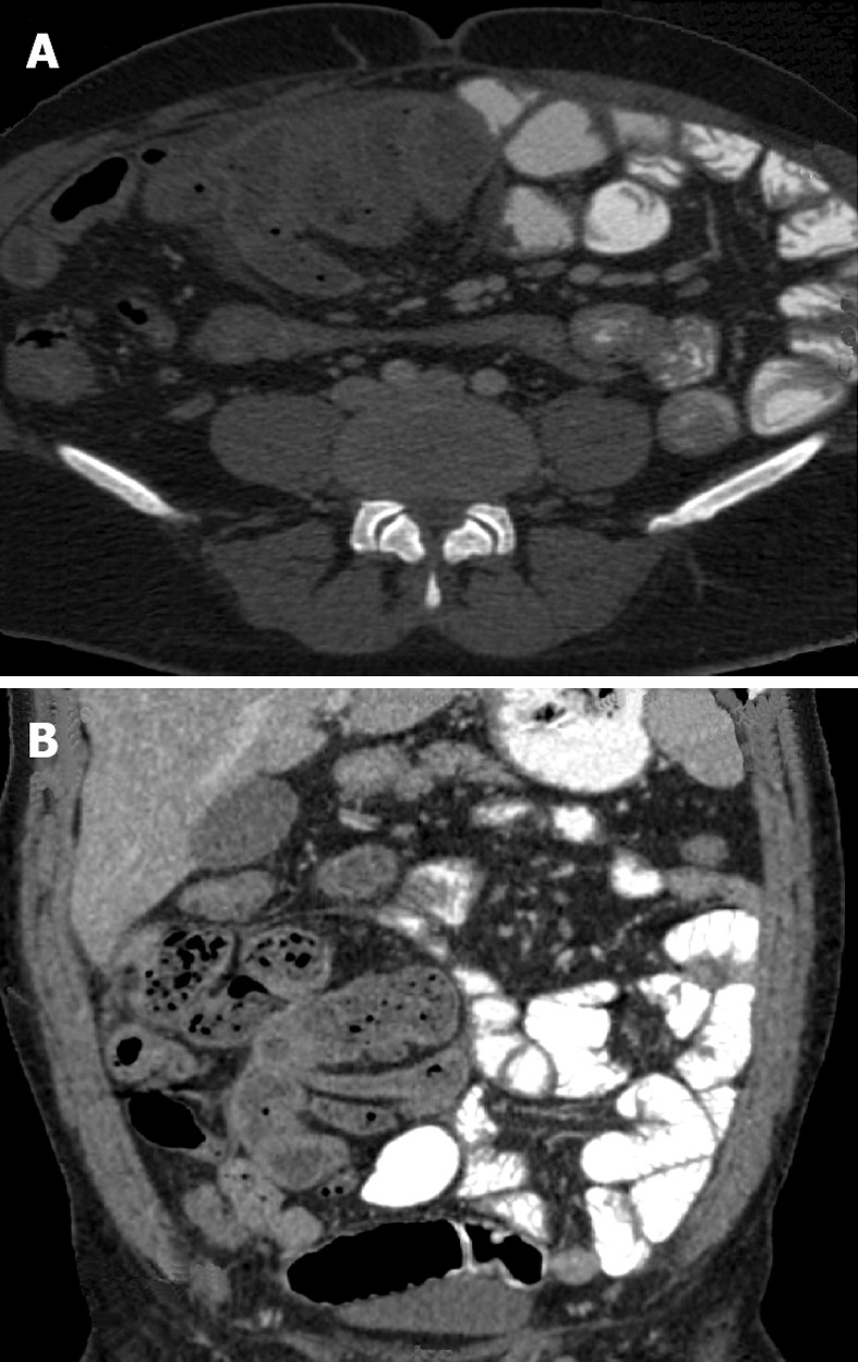
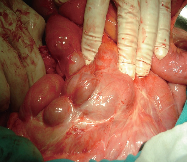
Abdominal cocoon, the idiopathic form of sclerosing encapsulating peritonitis, is a rare condition of unknown etiology that results in an intestinal obstruction due to total or partial encapsulation of the small bowel by a fibrocollagenous membrane. Preoperative diagnosis requires a high index of clinical suspicion. The early clinical features are nonspecific, are often not recognized and it is difficult to make a definite pre-operative diagnosis. Clinical suspicion may be generated by the recurrent episodes of small intestinal obstruction combined with relevant imaging findings and lack of other plausible etiologies. The radiological diagnosis of abdominal cocoon may now be confidently made on computed tomography scan. Surgery is important in the management of this disease. Careful dissection and excision of the thick sac with the release of the small intestine leads to complete recovery in the vast majority of cases.
Keywords: Computed tomography scan; Encapsulate; Intestinal obstruction; Peritonitis; Sclerosis; Surgery.
Figures
References
-
- Nakamoto H. Encapsulating peritoneal sclerosis--a clinician’s approach to diagnosis and medical treatment. Perit Dial Int. 2005;25 Suppl 4:S30–S38. - PubMed
-
- Wei B, Wei HB, Guo WP, Zheng ZH, Huang Y, Hu BG, Huang JL. Diagnosis and treatment of abdominal cocoon: a report of 24 cases. Am J Surg. 2009;198:348–353. - PubMed
-
- Mohanty D, Jain BK, Agrawal J, Gupta A, Agrawal V. Abdominal cocoon: clinical presentation, diagnosis, and management. J Gastrointest Surg. 2009;13:1160–1162. - PubMed
Publication types
MeSH terms
LinkOut - more resources
Full Text Sources
Medical
